Showing posts with label Medicine. Show all posts

Saturday, February 12, 2011

Fiber Tractography

Fiber tractography is the neurological procedure to study neuron axons tracts in a human brain using magnetic resonance imaging techniques and computer-based image analysis. A fiber tractography test can be shown in either two dimensional or three dimensional images. In the cerebrum the myelinated neuron axons of the cerebral cortex cluster together to form fibers, and these get together to form thick bundles or tracts called fasciculus which connect one lobe or region of the cortex with another. Thus, there is a complicated three-dimensional network formed by short connections among different cortical and subcortical regions.


The existence of these bundles has been revealed by histochemistry and biological techniques on post-mortem specimens. Brain tracts are not identifiable by direct exam, CT, or MRI scans. This difficulty explains the paucity of their description in neuroanatomy atlases and the poor understanding of their functions. The magnetic resonance imaging (MRI) sequences used look at the symmetry of brain water diffusion. Bundles of fiber tracts make the water diffuse asymmetrically in a tensor, the major axis parallel to the direction of the fibers. The asymmetry here is called anisotropy. There is a direct relationship between the number of fibers and the degree of anisotropy.

Thursday, February 10, 2011

Inferior Longitudinal fasciculus

The inferior longitudinal fasciculus is a bundle of myelinated axons which connects the occipital lobe with the temporal lobe in each hemisphere of the cerebrum. It runs parallel to the lateral walls of the inferior and posterior cornua of the lateral ventricle. Some anatomists call this bundle of fibers "occipitotemporal projection system." The function of the inferior longitudinal fasciculus is highly involved in visual memory as a 47-year-old woman with visual memory disturbance, demonstrated by the Wechsler Memory Scale-Revised, suffered from brain tumor in the right temporal lobe that disrupted the fibers bundle of the ILF. Disruption of white matter integrity in the inferior longitudinal fasciculus in teenagers with schizophrenia was also revealed by fiber tractography.

Wednesday, February 9, 2011

Superior Longitudinal Fasciculus

The superior longitudinal fasciculus is a thick and long bundle of myelinated axons which links the frontal lobe to the occipital, and part of the parietal and temporal lobes of each cerebral hemisphere. The association fibers that constitute the superior longitudinal fasciculus are bi-directional, which means that some axons originate in cortical neurons of the frontal lobe, while others in neurons located in the occipital and back regions of the temporal and parietal lobes, integrating motor and decision-making centers with visual and sensory ones. The superior longitudinal fasciculus sweeps along the superior margin of the claustrum in a great arc.

The superior longitudinal fasciculus consists of three distinct components: 1) SLF I is the dorsal component and originates in the superior and medial parietal cortex, passes around the cingulate sulcus and terminates in the dorsal and medial cortex of the frontal lobe and in the supplementary motor cortex; 2) SLF II is the major component of SLF and originates in the caudal-inferior parietal cortex and occipital lobe, ending in the dorsolateral prefrontal cortex (Brodmann 6, 8 and 46); 3) SLF III is the ventral component which begins in the supramarginal gyrus (rostral portion of the inferior parietal lobe) and ends in the ventral premotor and prefrontal cortex (Brodmann 6, 44, and 46).


Occipitofrontal Fasciculus

The occipitofrontal fasciculus is a bundle of association fibers which connects the frontal lobe with the occipital lobe of each brain hemisphere. The myelanated fibers that make up the occipitofrontal fasciculus radiate in a fan-like manner, extending into occipital and temporal lobes, lateral to the posterior and inferior cornua.


Tuesday, February 8, 2011

Uncinate Fasciculus

The uncinate fasciculus is a band of cortical neuron myelinated axons in the human brain that connects the inferior part of the frontal lobe with the anterior temporal lobe and parts of the limbic system, such as the hippocampus and amygdala that are located in this lobe. It is the last fasciculus to mature in the brain (beyond the age of 30). The average length of the uncinate fasciculus is 45 mm. It has three parts: a frontal extension, an intermediary segment, and a temporal segment. The uncinate fasciculus is a hook-shaped tract of fibers which go from the inferior frontal gyrus and the lower surfaces of the frontal lobe to the forward portions of the temporal lobe.

Uncinate Fasciculus and Schizophrenia

Evidence suggests that a disruption in connectivity between different brain regions, especially between the frontal and temporal lobes, linked up by the uncinate fasciculus, may partly explain some of the primary symptoms of schizophrenia. This idea, first proposed by Wernicke in 1906, posits a disturbance in functional connectivity between the frontal and temporal cortices in schizophrenia that might have as its basis a disruption in the white matter tracts connecting them. Abnormalities within the fiber bundles of the uncinate fasciculus associate with social anxiety, Alzheimer's disease, bipolar disorder, and depression in the elderly that had first had it in adolescence or early adulthood. Such abnormalities also link to schizophrenia.


Monday, February 7, 2011

Arcuate Fasciculus

The arcuate fasciculus is the thick bundle of myelinated axons which links the Broca's area to the Wernicke's area, which are the two language centers of the brain. Thus, the arcuate fasciculus fibers go from the inferior frontal gyrus of the frontal lobe to the back area of the superior temporal gyrus and a small area in the parietal lobe and vice versa. The function of this bundle of nerve fibers is to articulate or coordinate the motor verbal function of the Broca's area and the semantic and syntactic comprehension of the Wernicke's area. Damage to the arcuate fasciculus can cause a form of aphasia known as conduction aphasia, where auditory comprehension and speech articulation are preserved, but people find it difficult to repeat heard speech.


Sunday, February 6, 2011

Hypoglossal Nerve (CN XII)

Also known as CN XII, the hypoglossal nerve is the twelfth of twelve paired cranial nerves. The CN XII innervates the muscles of the tongue. It is a somatomotor nerve whose fibers originate in the hypoglossal nucleus neurons situated in the dorsal medulla oblongata of the brainstem. The hypoglossal nucleus nerve cells send axons that exit as rootlets that emerge in the ventrolateral sulcus of the medulla between the olive and pyramid. Then, the rootlets come together to make up the hypoglossal nerve, exiting the cranium through the hypoglossal canal.

As the CN XII comes out of the hypoglossal canal, it gives off a small meningeal branch, picking up a branch from the anterior ramus of C1. Then, it spirals behind the vagus nerve and passes between the internal carotid artery and internal jugular vein lying on the carotid sheath. After passing deep to the posterior belly of the digastric muscle, the hypoglossal nerve passes to the submandibular region to enter the tongue.


Saturday, February 5, 2011

Accessory Nerve (CN XI)

The accessory nerve is the eleventh of the twelve paired cranial nerves. It is also known as cranial nerve XI (CN XI). It is called accessory since it receives an accessory root from the upper part of the spinal cord as it emerges from the skull. The spinal fibers of the accessory nerve provides motor innervation from the central nervous system to two muscles of the neck: the sternocleidomastoid muscle and the trapezius muscle. The cranial part rapidly joins the vagus nerve and serves the same function as other vagal nerve fibers.

Although it originates in the central nervous system, the spinal accessory nerve begins outside the skull rather than inside, with its axonal fibers arising from neurons located in the upper spinal cord, near the medulla oblongata. These fibers coalesce to form the spinal accessory nerve, which enters the skull through the foramen magnum, the large opening at the base of the skull. Then the nerve runs along the inner wall of the skull towards the jugular foramen, through which it exits the skull together with the glossopharyngeal (CN IX) and vagus nerves (CN X). Thus, the accessory nerve is notable for being the only cranial nerve to both enter and exit the skull.


Friday, February 4, 2011

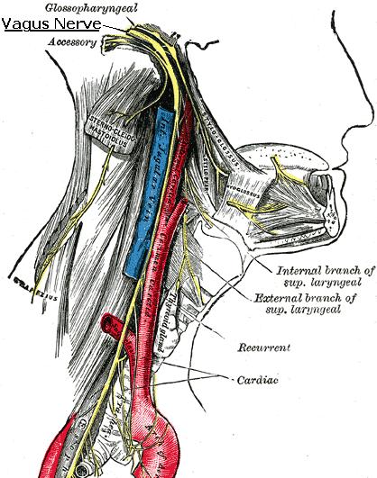
Vagus Nerve (CN X)

Also known as cranial nerve X (CN X), the vagus nerve is one of the twelve paired cranial nerves. Consisting of both motor and sensory fibers, the vagus nerve emerges from the medulla oblongata in the groove between the olive and the inferior peduncle. Then, it projects through the jugular foramen, passing into the carotid sheath between the internal carotid artery and the internal jugular vein down below the head, to the neck, chest and abdomen, where it contributes to the innervation of the viscera. Besides output to the various organs in the body the vagus nerve conveys sensory information about the state of the body's organs to the central nervous system. Between 80 and 90% of the fibers that make up the vagus nerve are sensory nerves communicating the state of the viscera to the brain.

The vagus nerve also supplies motor parasympathetic fibers to all the organs except the suprarenal glands, from the neck down to the second segment of the transverse colon. This means that the vagus nerve is responsible for such varied tasks as heart rate, gastrointestinal peristalsis, sweating, and quite a few muscle movements in the mouth, including speech (via the recurrent laryngeal nerve) and keeping the larynx open for breathing, via action of the posterior cricoarytenoid muscle, the only abductor of the vocal folds. It also has some afferent fibers that innervate the inner (canal) portion of the outer ear, via the Auricular branch, which also known as Alderman's nerve, and part of the meninges. This explains why a person may cough when tickled on their ear (such as when trying to remove ear wax with a cotton swab).


Wednesday, February 2, 2011

Vestibulocochlear Nerve (CN VIII)

The vestibulocochlear nerve, also known as cranial nerve VIII (CN VIII), is composed of sensory fibers which carry two types of sensory information to the brain: balance and hearing. It is the 8th of the twelve cranial nerves. The vestibulocochlear nerve is made up of bipolar neuron axons and splits into two branches: 1) the vestibular nerve, which carryies sensory information about balance; 2) the cochlear nerve, carrying information about hearing. The CN VIII enters the brain stem at the junction of the pons and medulla, lateral to the facial nerve. The auditory component of the eighth nerve terminates in a sensory nucleus called the cochlear nucleus, which is located at the junction of the pons and medulla. The vestibular part of the CN VIII ends in the vestibular nuclear complex located in the floor of the fourth ventricle.

Tuesday, February 1, 2011

Facial Nerve (CN VII)

The facial nerve is the seventh cranial nerve (CN VII). It is a paired nerve with both motor and sensory functions, providing motor innervation to the muscles of facial expression and sensory information of taste sensations from the anterior two-thirds of the tongue and oral cavity. It also supplies preganglionic parasympathetic fibers to several head and neck ganglia. The CN VII originates in the facial nerve nucleus in the pons while the sensory part of it arises from the nervus intermedius. It emerges from the brainstem between the pons and the medulla.

The motor fibers of the facial nerve innervate all the facial musculature; the principal muscles are the frontalis, orbicularis oculi, buccinator, orbicularis oris, platysma, the posterior belly of the digastric, and the stapedius muscle. The sensory fibers has the following components: 1) taste to the anterior two-thirds of the tongue; 2) secretory and vasomotor fibers to the lacrimal gland, the mucous membranes of the nose and mouth, and the submandibular and sublingual salivary glands; 3) cutaneous sensory impulses from the external auditory meatus and region back of the ear.


Monday, January 31, 2011

Abducens Nerve (CN VI)

The abducens nerve, also known as cranial nerve VI (CN VI), is one of the twelve paired cranial nerves. It is made up of motor axons and innervate the lateral rectus muscle of the eyeball. The CN VI originates in the abducens nucleus which is located in the pons, on the floor of the fourth ventricle. Motor axons leaving the abducens nucleus run ventrally and caudally through the pons, passing lateral to the corticospinal tract, which runs longitudinally through the pons at this level. Then, the abducens nerve leaves the brainstem at the junction of the pons and the medulla, medial to the facial nerve. In order to reach the eye, it runs upward, superiorly, and then bends forward, anteriorly.


Sunday, January 30, 2011

Trigeminal Nerve (CN V)

The trigeminal nerve, also known as cranial nerve V (CN V), is both a sensory and motor nerve which emerges from the side of the pons, near its upper border, by a large sensory and a small motor root. The sensory fibers of CN V carries sensory information of the head and face, and its motor fibers innervate (contract) the mastication muscles (biting and chewing). The cranial nerve V originates in the trigeminal nuclei located in the brainstem (pons, midbrain, medulla).

The name "trigeminal" means "three-branched", deriving from the fact that this paired nerve, one on each side of the pons, has three major branches: the ophthalmic nerve (CN V1), the maxillary nerve (CN V2), and the mandibular nerve (CN V3). The ophthalmic and maxillary nerves are purely sensory. The mandibular nerve has both sensory and motor functions. The three branches converge on the trigeminal ganglion, that is located within Meckel's cave, and contains the cell bodies of incoming sensory nerve fibers.


Saturday, January 29, 2011

Trochlear Nerve (CN IV)

The trochlear nerve, which is also called cranial nerve IV (CN IV), is the motor nerve which innervates the superior oblique muscle of the eyeball. The trochlear nerve is composed of axons of neurons situated in the trochlear nucleus, in the ventral part of the brainstem.

In terms of the number of axons that make it up, the cranial nerve IV is the smallest nerve of the twelve cranial nerves, but has the greatest intracranial length. Along with the optic nerve, it is the only cranial nerve that decussates (crosses to the other side) before innervating the superior oblique muscle. Trochlear nerve is also the only cranial nerve that exits from the dorsal aspect of the brainstem.


Friday, January 28, 2011

Oculomotor Nerve (CN III)

The oculomotor nerve, or cranial nerve III, is the third of twelve paired cranial nerves. It is responsible of eyelid and eyeball movement. The cranial nerve III (CN III) originates in the oculomotor nucleus situated in the superior colliculus of the midbrain.

When the oculomotor nerve emerges from the brain, it is invested with a sheath of pia mater, and enclosed in a prolongation from the arachnoid. It passes between the superior cerebellar (below) and posterior cerebral arteries (above), and then pierces the dura mater anterior and lateral to the posterior clinoid process, passing between the free and attached borders of the tentorium cerebelli.

The muscles that the CN III controls are the striated muscle in levator palpebrae superioris of the eyelid and all extraocular muscles of the eyeball, except for the superior oblique muscle and the lateral rectus muscle.


Thursday, January 27, 2011

Olfactory Nerve (CN I)

The olfactory nerve is one of the twelve cranial nerves that emerge directly from the base of the brain. Also called cranial nerve I (CN I), it is the first and shortest of the twelve. The olfactory nerve is formed by axons from specialized olfactory receptor neurons which are situated in the olfactory mucosa of the upper parts of the nasal cavity. These same nerve cells also projects axons that extend from the olfactory epithelium to the olfactory bulb, passing through the many openings of the cribriform plate of the ethmoid bone.

The nerve cells in the nasal cavity are chemosensitive and respond to chemical signals which are converted to electrical impulses that are carried up through the cranial nerve I to the olfactory bulb. The axons that make up the olfactory nerve continually regenerate throughout life as they project towards the olfactory bulb.


Tuesday, December 21, 2010

Retinal Ganglion Cells

A retinal ganglion cell (RGC) is a type of neuron (nerve cell), which is situated in the retina of the eye. Forming a thin layer, retinal ganglion cells receives visual information from photoreceptors (cones and rods) via two intermediate neuron types: bipolar cells and amacrine cells. The long axons of the retinal ganglion cells join together to make up the optic nerve and transmit image-forming and non-image forming visual information from the retina to several regions in the thalamus, hypothalamus, and mesencephalon, or midbrain.

In the human retina there are about 1.2 to 1.5 million retinal ganglion cells. With about 125 million photoreceptors per retina, on average each retinal ganglion cell receives inputs from about 100 rods and cones. Based on their projections and functions, there are at least five main classes of retinal ganglion cells: 1) midget cells, which project to the parvocellular layers of the lateral geniculate nucleus; 2) parasol cells; which project to the magnocellular layers of the lateral geniculate nucleus; 3) bistratified cells, which project to the koniocellular layers of the lateral geniculate nucleus; 4) photosensitive ganglion cells, which send their axons to the suprachiasmatic nucleus (SCN) via the retinohypothalamic tract for setting and maintaining circadian rhythms; 5) other ganglion cells extend their axons to the superior colliculus for eye movements (saccades).


Ocular Ischemic Syndrome

Ocular ischemic syndrome (OIS) refers to the ocular signs and symptoms which result from chronic vascular insufficiency (arterial hypoperfusion) to the eye. Severe ipsilateral or bilateral carotid artery stenosis is the most common cause of ocular ischemic syndrome. The syndrome has been associated with occlusion of the common carotid artery, internal carotid artery, and less frequently the external carotid artery. Other causes include: 1) severe ophthalmic artery occlusion, due to thromboembolism; 2) giant cell arteritis; 3) Takayasu's arteritis.

Common anterior segment signs of this eye condition include advanced cataract, anterior segment inflammation, and iris neovascularization. Posterior segment signs include narrowed retinal arteries, dilated but nontortuous retinal veins, midperipheral dot-and-blot retinal hemorrhages, cotton-wool spots, and optic nerve/retinal neovascularization. The presenting symptoms include ocular pain and abrupt or gradual visual loss.

Amaurosis fugax is a form of acute vision loss caused by reduced blood flow to the eye that may be a warning sign of an impending stroke. Consequently, those with transient blurring of vision are advised to urgently seek medical attention for a thorough evaluation of the carotid artery. Those with ocular ischemic syndrome are typically between the ages of 50 and 80; twice as many men than women are affected. More than 90% of those presenting with the condition have vision loss. Patients may report a dull, radiating ache over the eye and eyebrow. Those with ocular ischemic syndrome may also present with a history of other systemic diseases including arterial hypertension, diabetes mellitus, coronary artery disease, previous stroke, and hemodialysis.

Monday, December 20, 2010

Corneal Endothelium

The corneal endothelium is a single layer of specialized, flattened, mitochondria-rich cells that lines the posterior surface of the cornea and faces the anterior chamber of the eye. The corneal endothelium regulates fluid and solute transport across the posterior surface of the cornea and actively maintains the cornea in the slightly dehydrated state that is required for optical transparency.

The main physiological function of the corneal endothelium is to allow leakage of solutes and nutrients from the aqueous humor to the more superficial layers of the cornea while at the same time actively pumping water in the opposite direction, from the stroma to the aqueous.


Hexagonal cells of the corneal endothelium

Penetrating Keratoplasty

Penetrating keratoplasty is the surgical procedure in which a full thickness of the cornea is removed and replaced with donor tissue, using a microkeratome, which is an instrument employed for removing the cornea or a thin slice of it, creating a thin hinged flap. Penetrating keratoplasty is also used in the treatment of lattice corneal dystrophy. The aim of this eye surgery is to improve visual acuity by replacing the opaque host tissue by clear healthy donor tissue.

Penetrating Keratoplasty (Video)