Tuesday, June 30, 2009

Brainstem

The brainstem is the lower extension of the encephalon (brain). It is made up of the cerebral peduncles, the pons, and the medulla oblongata. The brainstem is structurally continuous with the spinal cord and provides the main motor and sensory innervation to the face and neck via the cranial nerves as it is the pathway for all fiber tracts passing up and down from peripheral nerves and spinal cord to the highest parts of the brain. The nerve connections of the motor and sensory systems from the main part of the brain to the rest of the body pass through the brainstem. This includes the corticospinal tract (motor), the posterior column-medial lemniscus pathway (fine touch, vibration sensation and proprioception) and the spinothalamic tract (pain, temperature, itch and crude touch). The brainstem controls vital functions such as breathing and instructing the heart to beat.

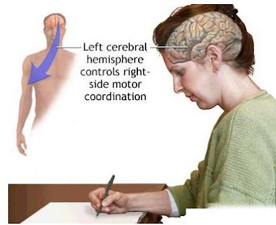
All information related from the body to the cerebrum and cerebellum and vice versa, must go through the brainstem. The ascending pathways that come from the body to the brain are the sensory pathways, and include the spinothalamic tract for pain and temperature sensation and the dorsal column, fasciculus gracilis, and cuneatus for touch, proprioception, and pressure sensation (both of the body). (The facial sensations have similar pathways, and will travel in the spinothalamic tract and the medial lemniscus also). Descending tracts are upper motor neurons destined to synapse on lower motor neurons in the ventral horn and intermediate horn of the spinal cord. In addition, there are upper motor neurons that originate in the brain stem's vestibular, red, tactile, and reticular nuclei, which also descend and synapse in the spinal cord.


Monday, June 29, 2009

Medulla Oblongata

The medulla oblongata is the lower portion of the brainstem. Lying below the pons and and anterior to the cerebellum, the medulla oblongata looks like a swelling at the top of the spinal cord. It controls autonomic functions, such as breathing and blood pressure, and relays nerve signals between the cerebral cortex and spinal cord. The cardiac center is located in the medulla oblongata and is responsible for controlling the heart rate.

The anterior portion of the medulla oblongata is called the pyramid and lies between the anterior median fissure and the antero-lateral sulcus. The medulla controls the reflex actions of the pharynx, larynx, and tongue, which are related to deglutition, mastication, and speech, as well as the visceral reflexes of coughing, sneezing, sucking, vomiting, and salivating, and other secretory functions.

Saturday, June 27, 2009

Pons

The pons is a structure which makes up the upper segment of the brain stem. Caudal to the midbrain and cranial to the medulla oblongata, the pons contains nerve fibers that connect the two halves of the cerebellum. The pons plays an important role in coordinating movements involving right and left sides of the body as it relays sensory information between the cerebellum and the cerebral cortex, controling arousal, and regulating respiration. Below the pons is the medulla, which is continuous with the spinal cord and transmits ascending and descending nerve fibers between the spinal cord and the brain. The pons is irrigated by pontine arteries.

Friday, June 26, 2009

Cerebral Peduncles

The cerebral peduncles are two large bands of white matter composed of axons which descends from upper motor neurons in the frontal lobes of the cerebral cortex which emerge from the underside of the cerebral hemispheres. The cerebral peduncles approach each other as they enter the rostral border of the pons. The important fibers running through the cerebral peduncles include the corticospinal tract and the corticobulbar tract, among others.


Thursday, June 25, 2009

Midbrain

The midbrain is a small region of brain situated between the diencephalon and the pons. It contains a number of tracts of nerve fibers, which include the pyramidal tract, involved in the performance of motor skills. The midbrain also contains the nerve pathways between the cerebral hemispheres and the medulla oblongata, and the nuclei of the third and fourth cranial nerves.

The midbrain comprises the tectum, tegmentum, the ventricular mesocoelia, and the cerebral peduncles. Caudally the midbrain adjoins the pons, and rostrally it adjoins the thalamus and hypothalamus. The midbrain, or mesencephalon, develops from the middle section of the embryonic brain. The human midbrain is archipallian in origin, which means its general architecture is shared with the most ancient of vertebrates. Dopamine produced in the substantia nigra plays a role in motivation and habituation of species.

Wednesday, June 24, 2009

Lobotomy

Also known as a leukotomy, lobotomy is a neurosurgical procedure which consists of cutting the connections to and from the prefrontal cortex of the cerebrum. Psychiatrists and neurosurgeons no longer perform lobotomies on patients. Now they use various drugs, such as antipsychotic chlorpromazine (Thorazine), and psychological therapies to treat mental health issues. Lobotomies were used mainly from the 1930s to 1950s to treat a wide range of severe mental illnesses, including schizophrenia, clinical depression, and various anxiety disorders. The patient's informed consent in the modern sense was often not obtained.

Lobotomy is a surgical incision into the frontal lobe of the brain to sever one or more nerve tracts, a technique formerly used to treat certain mental disorders but now rarely performed. In 1935, Portuguese physician and neurologist António Egas Moniz pioneered a surgery he called prefrontal leucotomy. The procedure involved drilling holes in the patient's head and destroying tissue in the frontal lobes by injecting alcohol. He later changed technique, using a surgical instrument called a leucotome that cut brain tissue by rotating a retractable wire loop. Moniz was given the Nobel Prize for medicine in 1949 for this work.

The American neurologist and psychiatrist Walter Freeman was intrigued by Moniz's work, and with the help of his close friend, a neurosurgeon named James W. Watts, he performed the first prefrontal leucotomy in the U.S. in 1936. Freeman and Watts gradually refined the surgical technique, and created the Freeman-Watts procedure (the "precision method," the standard prefrontal lobotomy).

The Freeman-Watts prefrontal lobotomy still required drilling holes in the scalp, so surgery had to be performed in an operating room by trained neurosurgeons. Walter Freeman believed this surgery would be unavailable to those he saw as needing it most: patients in state mental hospitals having no operating rooms, surgeons, or anesthesia, and limited budgets. Freeman wanted to simplify the procedure so that it could be carried out by psychiatrists in mental asylums, which housed roughly 600,000 American inpatients at the time.

Phineas Gage case was an accidental lobotomy in which Phineas became almost childish in his behavior, unwilling to listen to others and often using obscenities. The part of Phineas' brain that had been destroyed by this accidental lobotomy was the orbitofrontal cortex in situated in the frontal lobe.

Tuesday, June 23, 2009

Phineas Gage Case (Frontal Lobe Damage)

In September, 1848, Phineas Gage, foreman of a road construction gang, had an iron rod blown through his head, achieving immortality. But Phineas Gage did not become immortal in the usual way by going directly to his heavenly reward, for he survived. In fact, it was the details of his survival which constituted the basis for the very considerable amount of fame that came his way.

It seems that Phineas Gage had poured a charge of powder into a hole in a rock, prior to a routine blasting operation. The usual procedure was then for an assistant to cover the powder with sand. For some reason this had not been done, and Gage neglected to check on the matter. Instead, supposing the sand covering to be in place, he dropped a heavy tamping iron into the hole. The result was catastrophic: the iron rod struck upon rock, made a spark, ignited the powder inside the hole, and took off for the stratosphere. On its way the rod, which was nearly 4 feet long, passed cleanly through Phineas' brain, entering high in his left cheek and coming out from the top of his head, causing frontal lobe damage.

Phineas Gage was stunned for an hour, after which, with some assistance, he was able to walk off to see a surgeon, talking on the way about the hole in his head. Eventually he recovered from the infection that developed in the wound and lived for another twelve years. Scientists verified the story by actual examination of the damaged brain. It was found that not only the left frontal lobe had been severely damaged, but the damage had spread to the right frontal lobe as well.

Gage's surprising survival of such a spectacular injury was followed by equally surprising aftereffects. The aftereffects were remarkable precisely because of their nonspectacular nature. For Gage could still displayed no loss of memory and he was still able to perform his job. For a man with such extensive damage of the very portion of the cerebrum which had long been believed to be the seat of the higher intellectual processes, Gage displayed a disproportionately small decrease in his mental capacities. Yet, there were some changes in Phineas Gage, though, but they were of quite a different nature from what what would have been predicted by the then prevailing theories.

It seemed that his personality rather than his memory had been mainly affected. Before the accident Phineas Gage had been considerate, efficient, and well-balanced. Afterward he was fitful and irreverent, indulging frequently in gross profanity and manifesting little consideration for others. He had become obstinate yet capricious and vacillating. With these new traits Gage could no longer be trusted to supervise others. In fact, he showed little inclination toward work of any kind, but instead chose to travel around, making a living by exhibiting himself and his tamping iron.

Monday, June 22, 2009

Motor Strip

The motor strip is a long narrow area in the frontal lobe. It is situated in the anterior central gyrus and runs down parallel to and in front of the central sulcus. The motor strip controls all bodily movements; the motor strip in the frontal lobe of the left cerebral hemisphere controls the movement of the right side of the body; and that of the right controls the movement of the left side of the body.

The most striking aspect of the motor strip map is that the areas assigned to various body parts on the cortex are proportional not to their size, but rather to the complexity of the movements that they can perform. Hence, the areas for the hand and face are especially large compared with those for the rest of the body. This is no surprise, because the speed and dexterity of human hand and mouth movements are precisely what give us two of our most distinctly human faculties: the ability to use tools and the ability to speak.

Sunday, June 21, 2009

Parietal Lobe

The parietal lobe is the central portion of the cerebral cortex, situated between the frontal lobe and occipital lobe, above the temporal lobe. The central salcus separates the parietal lobe from the frontal lobe, and the parieto-occipital sulcus partially divides it from the occipital lobe.

The parietal lobe contains the posterior central gyrus, the superior parietal lobule, the inferior parietal lobule, and parieto-occipital lobule. The parietal lobe integrates sensory information from various parts of the body, as well as knowledge of numbers and their relations, and is involved in the manipulation of objects. Portions of the parietal lobe are involved with visuospatial processing. Although multisensory in nature, the posterior parietal cortex is often referred to by vision scientists as the dorsal stream of vision (as opposed to the ventral stream in the temporal lobe). This dorsal stream has been called both the 'where' stream, as in spatial vision, and the 'how' stream, as in vision for action.

Friday, June 19, 2009

Primary Auditory Cortex

The primary auditory cortex is the area of the cerebrum which is involved in processing auditory (sound) information. The primary auditory cortex is situated in the temporal lobe, in the posterior half of the superior temporal gyrus, just below the lateral sulcus. Auditory signals reach perception only if received and processed by that particular cortical area of the temporal lobe.

Neurons in the auditory cortex are organized according to the frequency of sound to which they respond best. Neurons at one end of the auditory cortex respond best to low frequencies, while neurons at the other respond best to high frequencies. Brief auditory stimuli activate the primary auditory cortex earlier than any other cortical area so, within a certain latency range, the primary auditory cortex is the only cortical source contributing to the auditory evoked field.

Thursday, June 18, 2009

Temporal Lobe

The temporal lobe is an of the cerebral cortex which is situated beneath the lateral sulcus on both the left and right hemispheres of the brain. The temporal lobe is associated with auditory processing and is home to the primary auditory cortex. It also processes semantics in both speech and vision. The temporal lobe contains the hippocampus and plays a key role in the formation of long-term memory.

Auditory signals from the cochlea, which are relayed through several subcortical nuclei, reach the temporal lobe at the superior temporal gyrus within the lateral sulcus. This part of the temporal lobe, which is called the primary auditory cortex, is involved in hearing. Adjacent areas in the superior, posterior and lateral parts of the temporal lobes participate in high-level auditory processing. Wernicke's area, which spans the region between temporal and parietal lobes, plays a key role in speech, in tandem with Broca's area, which is in the frontal lobe. The functions of the left temporal lobe are not limited to low-level perception but extend to comprehension, naming, verbal memory and other language functions. Sound processing is controlled by the temporal lobes- in the Broca’s area and Wernicke’s area.

Wednesday, June 17, 2009

Central Sulcus

The central sulcus is a fissure in the cerebral cortex of each hemisphere. It is also called the fissure of Rolando or the Rolandic fissure, after the anatomist Luigi Rolando. The central sulcus separates the parietal lobe from the frontal lobe and the primary motor cortex from the primary somatosensory cortex.

The motor cortex is on one side of the central sulcus; the sensory cortex is on the other (in the parietal lobe). The central sulcus has a 'map' of the human body on each side that corresponds to the other side. When the sensory part is stimulated, its associated motor part is right across the sulcus.

Tuesday, June 16, 2009

Lateral Sulcus

The lateral sulcus, also called Sylvian fissure, is the deepest cortical fissure of the brain. It stretches between the frontal and temporal lobes, then goes back and upward over the lateral aspect of the cerebral hemisphere. The lateral sulcus divides the frontal lobe and parietal lobe above from the temporal lobe below. The lateral sulcus is situated in both cerebral hemispheres but it is longer in the left hemisphere.

The Sylvian fissure has a number of side branches. Two of the most prominent and most regularly found are the ascending ramus and the horizontal ramus of the lateral fissure, which subdivide the inferior frontal gyrus. The lateral sulcus also contains the transverse temporal gyri, which are part of the primary auditory cortex.


Amygdala

The amygdala is an almond shaped subcortical structure located deep within the temporal lobes, medial to the hypothalamus and adjacent to the hippocampus. The amygdala has long been linked with a person's mental and emotional state. The regions described as amygdalae comprises several nuclei with distinct functional traits. Among these nuclei are the basolateral complex, the centromedial nucleus and the cortical nucleus. The basolateral complex can be further subdivided into the lateral, the basal and the accessory basal nuclei.

The amygdalae is connected to the hypothalamus, to the thalamic reticular nucleus for increased reflexes, to the nuclei of the trigeminal nerve and facial nerve which implements facial muscle movements, and to the ventral tegmental area, locus coeruleus, and laterodorsal tegmental nucleus for activation of dopamine, norepinephrine and epinephrine. The amygdala plays a critical role in the expression of emotions and the learning of new emotional responses. Much evidence suggests that human anxiety disorders result from anomalies in amygdala function.

Phylogenetically, the amygdala is a very old structure; probably very early on in Phylogeny. It was primarily involved in protecting organisms, moving them away from obnoxious chemical milieu. As organisms evolved the amygdala got different kinds of sensory information in to evaluate stimuli in the environment, and that is one of the reasons why it is more highly connected with the neocortex as organisms evolved. It's getting more and more high-level information to do an interpretation of what is going on in the environment.

Monday, June 15, 2009

Cortical Magnification Factor

The cortical magnification factor is the apportioning of proportionally more space on the cortex to the representation of specific areas of sensory receptors. For example, a small area on the retina in or near the fovea receives more space on the cortex than the same area of peripheral retina. Smilarly, the fingertips receive more space on the somatosensory cortex than the forearm or leg. The cortical magnification factor is normally expressed in millimeters of cortical surface per degree of visual angle. When expressed in this way, the values of cortical magnification factor vary by a factor of approximately 100 between the foveal and peripheral representation of the primary visual cortex (V1) of primates.

Cortical magnification describes how many neurons in an area of the visual cortex are 'responsible' for processing a stimulus of a given size, as a function of visual field location. In the center of the visual field, corresponding to the fovea of the retina, a very large number of neurons process information from a small region of the visual field. If the same stimulus is seen in the periphery of the visual field, it would be processed by a much smaller number of neurons. The reduction of the number of neurons per visual field area is achieved in several steps along the visual pathway, starting already in the retina.

Lateral Geniculate Nucleus

The lateral geniculate nucleus (LGN) is a subcortical structure which lies in each cerebral hemisphere. It is the primary processing center for visual information received from the retina of the eye. The lateral geniculate nucleus is located inside the thalamus of the brain and is thus part of the central nervous system. The lateral geniculate nucleus consists of six layers of neurons with each alternating layer receiving inputs from a different eye: 3 layers for the left eye and 3 layers for the right.

The lateral geniculate nucleus receives information directly from the ascending retinal ganglion cells via the optic nerve and from the reticular activating system. Neurons of the lateral geniculate nucleus send their axons through the optic radiations, a pathway directly to the primary visual cortex, also known as the striate cortex. The primary visual cortex surrounds the calcarine fissure, a horizontal fissure in the medial and posterior occipital lobe. In addition, the lateral geniculate nucleus gets many strong feedback connections from the primary visual cortex.

Two types of information: motion vs. color and form, are kept in separate layers in the lateral geniculate nucleus; these being the magnocellular and parvocellular layers, respectively. The lateral geniculate nucleus is innervated by most of the optic tract axons arising from retinal ganglion cells. In turn, neurons in the lateral geniculate nucleus give rise to axons that project by way of the optic radiation to the striate cortex in the ipsilateral occipital lobe.


Ventral Stream

The ventral stream is the area of the cerebral cortex which runs from the occipital lobe to the temporal lobe. The ventral stream is connected to the medial temporal lobe, which stores long-term memories, the limbic system, and the dorsal stream, which deals with object locations and motion. It is responsible for visual perception and object recognition. The ventral stream receives its main input from the parvocellular layer of the lateral geniculate nucleus of the thalamus. These neurons project to V1 sublayers 4Cß, 4A, 3B and 2/3a successively.

Sunday, June 14, 2009

Dorsal Stream

The dorsal stream is the cerebral cortex area which begins in the occipital lobe, where the primary visual cortex is located, and ends in the parietal lobe. The dorsal stream processes spatial information as it is involved in spatial awareness, recognizing where objects are in space, and guidance of actions. The dorsal stream is essential for the perception and interpretation of spatial relationships, accurate body image, and the learning of tasks involving coordination of the body in space. It is interconnected with the parallel ventral stream which runs downward from V1 into the temporal lobe.

Saturday, June 13, 2009

Optic Tract

The optic tract is the extension of the visual system pathway that stretches from the optic chiasm to the lateral geniculate nucleus of the thalamus. The optic tract is a continuation of the optic nerves that cross over at the optic chiasm. Each optic tract contains axons from ganglion cells in the retinas of both the left and right eyes, but information from only one half of each eye's visual field.

Friday, June 12, 2009

Primary Visual Cortex

The primary visual cortex is situated in the occipital lobe of the brain, in and around the calcarine fissure. It is anatomically equivalent to Brodmann area 17. The primary visual cortex receives information from the lateral geniculate nucleus and transmits information to two primary pathways, which are called the dorsal stream and the ventral stream.

Neurons in the primary visual cortex fire action potentials when visual stimuli appear within their receptive field. But for any given neuron, it may respond to a subset of stimuli within its receptive field. This property is called neuronal tuning. The visual cortex receives its blood supply primarily from the calcarine branch of the posterior cerebral artery.

Optic Chiasm

The optic chiasm is the point at which the optic nerves cross over. The optic chiasm is situated at the bottom of the cerebrum immediately below the hypothalamus. Nervous impulses from retinal cells follows the optic nerve to the optic chiasm, where half the fibers from each eye cross to the opposite cerebral hemisphere. Visual information from the right half of each retina travels to the right occipital lobe, and visual information from the left half of each retina travels to the left occipital lobe.


Thursday, June 11, 2009

Olfactory Bulb

The olfactory bulb is the part of the vertebrate forebrain that is involved in olfaction, that is to say the perception of odors. In humans, the olfactory bulb is on the inferior and bottom side of the brain. The olfactory bulb is supported and protected by the cribriform plate, which separates it from the olfactory epithelium, and which is perforated by olfactory neurons axons.

The olfactory bulb is divided into two different structures: the main olfactory bulb, and the accessory olfactory bulb. The main olfactory bulb has a multi-layered cellular architecture made up of the glomerular layer, external plexiform layer, mitral cell layer,internal plexiform layer, granule cell layer. the glomerular layer receives direct input from olfactory nerves, which consist of axons from approximately ten million olfactory receptor neurons in the olfactory mucosa, a region of the nasal cavity.

Archicortex

The archicortex is a portion of the cerebral cortex that, together with the paleocortex, develops in association with the olfactory system. The archicortex is phylogenetically older than the neocortex and lacks its layered structure. The embryonic archicortex corresponds to the cortex of the dentate gyrus and hippocampus in mature mammals. It is part of the limbic system and has functions related to emotions and formation of memory. Signals being sent from the limbic lobe to the hippocampal formations go via the archicortex as an intermediate.

Wednesday, June 10, 2009

Paleocortex

The paleocortex is the portion of the cerebral cortex, intermediate phylogenetically between the neocortex and archicortex, and which develops in association with the olfactory system. The paleocortex is less stratified than the neocortex and consists chiefly of the piriform cortex and the parahippocampal gyrus.

Tuesday, June 9, 2009

Allocortex

The allocortex is a part of the cerebral cortex which have fewer neurons layers (fewer than six) than the isocortex, or neocortex. The regions of the cerebrum that are described as part of the allocortex are: 1) the olfactory cortex, which mediates olfaction; 2) the hippocampus, which mediates limbic input.

The allocortex is also referred to as the "heterotypic cortex." Phylogenetically, the allocortex is the oldest part of the cerebral cortex.

Monday, June 8, 2009

Cerebral Lateralization

Cerebral lateralization is a relatively new area of study which has important implications for the analysis of the relationship between cognition and emotion. The study of the functions of the left hemisphere and the right hemisphere suggests that there are different kinds of cognition, that is to say, different ways of knowing reality.

The research of cerebral lateralization has dug up evidence that each of the cerebral hemispheres is associated with different kinds of cognition. The left hemisphere is analytic, breaking sense data into meaningful pieces which are equivalent to one another; it is also linear and tend to organize these pieces into sequences, and symbolic, in that it is adept to attaching sense data to learned shapes and sounds. These abilities explain the great importance of the left hemisphere in the control of language. In most people, when the left hemisphere is damaged in certain places, crippling desorders of language result, such as aphasias.

Cerebral lateralization of the right hemisphere, in contrast, is associated with a wholistic, synthetic sort of cognition, which has been termed syncretic cognition (Tucker, 1981). Tucker suggests that this provides an integration of sensory information from different channels, such as visceral, visual, auditory, tactual, etc, into a superordinate conceptualization. There is much recent evidence which suggests that the right hemisphere plays a special role in both the expression and recognition of emotion. Input of the subcortical and limbic system mechanisms associated to higher brain structures is lateralized. Thus important fiber systems associated with motivation and emotion, such as the medial forebain bundle, are right-lateralized.

The left side of the brain has abilities and attributes which could be likened to the functioning of a computer, as it receives, stores and processes information fed into it from external sources. Its working out methods are primarily of a binary either/or, yes/no, accept/reject nature and are strictly logical in the sense that they use the in-coming information in an ordered, predictable manner within definite, limited parameters. The left hemisphere function could be described as passive in that its operation is predominantly reactive. It can never generate from itself any process or task for which it does not have already introduced, or memorized, data.

Through the observation of cerebral lateralization it has been found that the left hemisphere feels secure in the continuity of a process. It does not question how the inception of the process came about, or whether the continuation of it is still valid, and does not care to contemplate the ending of it. The stability of the indefinite continuing is sufficient in itself so that the left will tend to fear, and hence resist, any threat of discontinuity. However, the right hemisphere is the vehicle for change, as it is the medium for such phenomena as creative thinking, initiatives, enterprise and inspiration. In fact, without the left having been conscious of it, the right will have been responsible for initiating many of the processes which the left then tenaciously continues.

In a mature and healthy human being, the right side will initiate reasonable and beneficial processes, will lend support to valid and proven processes, and, with resolution, terminate those which have passed their usefulness or, even worse, have become definitely harmful to the physical and mental well-being.

Whereas the left likes to define, categorize, and concretize the particular as separate, definite and contained, the right seeks always to synthesize and integrate, since it regards relationship not as evidence of multiplicity, but over-riding unity. It will not be satisfied with anything less than the whole meaning.

Saturday, June 6, 2009

Right Cerebral Hemisphere

The right cerebral hemisphere is one of the two longitudinal halves into which the cerebrum is divided. It is divided into four lobes: the frontal, temporal, parietal, and occipital. The right hemisphere motor strip controls the muscular movement of the left side of the body; that is to say that left-handed people are lateralized towards the right hemisphere.

The right cerebral hemisphere is connected to the left hemisphere through the corpus callosum, which is a band of white matter composed of myelinated fibers. The right hemisphere is linked to the basal ganglia and thalamus through myelinated axons which go from its frontal lobe to these regions.

The right cerebral hemisphere functions differently from the left, as it seems to have a complete different way of processing the data, or different approach to reality. It is wholistic, which means that the right hemisphere does not analyze incoming information, breaking it into parts, but synthesize it into one indivisible whole. It is also intuitive and imaginative.


Friday, June 5, 2009

Left Cerebral Hemisphere

The left cerebral hemisphere is one of the two longitudinal cerebral halves of the human brain. Like its opposite, it is divided into four lobes; frontal, temporal, parietal, and occipital. The left cerebral hemisphere is connected to the right hemisphere through the corpus callosum.

The left cerebral hemisphere motor strip controls movement of the right side of the body. Depending on the severity, a stroke affecting the left cerebral hemisphere may result in functional loss or motor skill impairment of the right side of the body, and may also cause loss of speech.

Linear and analytical reasoning and language skills such as oral articulation of words and grammar comprehension are often lateralized to the left hemisphere of the brain, which contains the Broca's cortical area and the Wernicke's. Dyscalculia is a neurological syndrome associated with damage to the left temporo-parietal junction.

Thursday, June 4, 2009

Cerebral Hemispheres

The cerebral hemispheres are the two longitudinal halves into which the cerebrum is divided. The cerebrum (brain) is divided into two hemispheres; a left cerebral hemisphere and a right cerebral hemisphere. These two hemispheres are joined together by the corpus callosum, which is a thick band of white matter made up of myelinated axons connecting the left hemisphere cortex to the right hemisphere cortex. Each cerebral hemisphere takes care of one side of the body, but the motor controls are crossed: the right hemisphere controls the left side of the body, whereas the left hemisphere controls the muscular contractions of the right side of the body.

Although the cerebral hemispheres are very similar in appearance, they differ in their function in most people. The left hemisphere is analytical, logical, and linear, whereas the right is wholistic, intuitive, creative and imaginative. But the most concrete evidence of hemispheric lateralization for one specific ability is language. The major areas involved in language skills, Broca's area and Wernicke's area, are in the left hemisphere. Perceptual information from the eyes, ears, and rest of the body is sent to the opposite hemisphere, as motor impulses are sent out to the opposite side of the body; this is due to the crossing over of motor fibers at the pyramid of the medulla oblongata.


Wednesday, June 3, 2009

Dorsolateral Prefrontal Cortex

The dorsolateral prefrontal cortex is located between the superior frontal gyrus and the inferior frontal gyrus of the frontal lobe, covering the middle central gyrus and straddling the anterior halves of the superior frontal sulcus and the inferior frontal sulcus. The dorsolateral prefrontal cortex is the last area to develop (myelinate) in the human cerebrum. This area mainly receives its blood supply from the middle cerebral artery. With respect to neurotransmitter systems, there is evidence that dopamine plays a particularly important role in the dorsolateral prefrontal cortex.

The dorsolateral prefrontal cortex is connected to the orbitofrontal cortex, and to a variety of brain areas, which include the thalamus, parts of the basal ganglia, the hippocampus, and primary and secondary association areas of neocortex, including posterior temporal, parietal, and occipital lobes.

The dorsolateral prefrontal cortex plays an important role in the integration of sensory and mnemonic information and the regulation of intellectual function and action. It is the highest cortical area responsible for organization, motor planning, and regulation. It is also involved in working memory. Nevertheless, the dorsolateral prefrontal cortex is not exclusively responsible for the executive functions. The complex human mental activity require the additional cortical and subcortical circuits that the dorsolateral prefrontal cortex is connected with.

Tuesday, June 2, 2009

Orbitofrontal Cortex

The orbitofrontal cortex is the foremost part of the frontal lobe, lying on the roof of the orbit. The orbitofrontal cortex is involved in cognitive processes such as decision-making. Considerable individual variability has been found in the orbitofrontal cortex of both humans and non-human primates. Because of its functions in emotion and reward, the orbitofrontal cortex is considered by some to be a part of the limbic system. It has been proposed that the orbitofrontal cortex is involved in sensory integration, in representing the affective value of reinforcers, and in decision-making and expectation. It is thought to regulate planning behavior associated with sensitivity to reward and punishment.

Although the orbitofrontal cortex is one of the least explored and least understood regions of the human cerebral cortex, clinical evidence suggests that the orbitofrontal cortex is involved in critical human functions, such as social adjustment and the control of mood, drive and responsibility, traits that are crucial in defining the ‘personality’ of an individual. Phineas Gage is a paradigmatic patient, who, after suffering major destruction of the orbital and medial prefrontal cortices in both hemispheres, was portrayed as no longer being himself.

Monday, June 1, 2009

Visual Center of the Brain

The visual center of the brain is located in the occipital lobe of the cerebral cortex. The visual center comprises the primary visual cortex, which is Brodmann area 17, commonly called V1, of both cerebral hemispheres.

The visual center of the brain receives and processes the images from the eye retina that arrives via the optic nerves and optic chiasma. It projects these images into parietal and temporal areas of the cortex known as dorsal stream and ventral stream respectively.

Calcarine Fissure

The calcarine fissure is the cerebral fold which is situated in the occipital lobe of the brain. It is also called calcarine sulcus. The calcarine sulcus begins near the occipital pole and runs up forward to a point just below the splenium of the corpus callosum, where it is joined at an acute angle by the medial part of the parietooccipital fissure.

The calcarine fissure is a part of the primary visual cortex, where the visual center of the brain is located. The central visual field is located in posterior portion of the calcarine sulcus and the peripheral visual field in the anterior portion. The amount of cortex dedicated to each square millimeter of the visual field is highly non-proportional, which means that significantly more cortex is dedicated to the processing of information originating from the fovea than other locations.