Neomycin is a common antibiotic which belongs the aminoglycoside group of antibiotics. It is found in many topical medications such as ointments, eyedrops, and cream. Neomycin is a broad spectrum antibiotic effective against both gram positive and gram negative bacteria, interfering with protein synthesis in sensitive bacterial cells such as species of Proteus and Staphylococcus.
Neomycin is commonly used as a topical preparation like Neosporin. It can be given orally, too, but it is usually combined with other antibiotics. Neomycin is not absorbed from the gastrointestinal tract, and has been used as a preventative measure for hepatic encephalopathy and hypercholesterolemia.
Neomycin was discovered by the microbiologist Selman Waksman and his student Hubert Lechevalier at Rutgers University in 1949. It is produced naturally by the bacterium Streptomyces fradiae.
Thursday, February 26, 2009
Monday, February 23, 2009
Aminoglycosides
An aminoglycoside is a molecule composed of a sugar group and an amino group. Several aminoglycosides function as antibiotics that are effective against certain types of bacteria. They include amikacin, arbekacin, gentamicin, kanamycin, neomycin, netilmicin, paromomycin, rhodostreptomycin, streptomycin, tobramycin, and apramycin. Anthracyclines are another group of aminoglycosides. These compounds are used in chemotherapy.
Aminoglycosides are potent bactericidal antibiotics that act by creating fissures in the outer membrane of the bacterial cell. They are particularly active against aerobic, gram-negative bacteria and act synergistically against certain gram-positive organisms. Amikacin may be particularly effective against resistant organisms. Aminoglycosides that are derived from bacteria of the Streptomyces genus are named with the suffix -mycin, while those which are derived from Micromonospora are named with the suffix -micin.
Aminoglycosides are used in the treatment of severe infections of the abdomen and urinary tract, as well as bacteremia and endocarditis. They are also used for prophylaxis, especially against endocarditis. Resistance is rare but increasing in frequency. Avoiding prolonged use, volume depletion and concomitant administration of other potentially nephrotoxic agents decreases the risk of toxicity.
The first aminoglycoside, streptomycin, was isolated from Streptomyces griseus in 1943. Neomycin, isolated from Streptomyces fradiae, had better activity than streptomycin against aerobic gram-negative bacilli but, because of its formidable toxicity, could not safely be used systemically. Gentamicin, isolated from Micromonospora in 1963, was a breakthrough in the treatment of gram-negative bacillary infections, including those caused by Pseudomonas aeruginosa. Other aminoglycosides were subsequently developed, including amikacin (Amikin), netilmicin (Netromycin) and tobramycin (Nebcin), which are all currently available for systemic use in the United States.
Saturday, February 21, 2009
Gentamicin
Gentamicin is a water-soluble antibiotic which belongs to the aminoglycoside group and is used to treat many types of bacterial infections, particularly those caused by Gram-negative bacteria. However, gentamicin is not used for Neisseria gonorrhoeae, Neisseria meningitidis or Legionella pneumophila bacterial infections. It is synthesized by Micromonospora, a genus of Gram-positive bacteria widely present in the environment (water and soil). To highlight their specific biological origins, gentamicin and other related antibiotics produced by this genus have generally their spellings ending in ~micin and not in ~mycin.
Gentamicin is a bactericidal antibiotic that works by binding the 30S subunit of the bacterial ribosome, interrupting protein synthesis. It is administered intravenously, intramuscularly or topically to treat infections. Gentamincin is a powerful antibiotic which can destroy a wide range of bacteria, normally Gram-negative bacteria, such as Pseudomonas, Proteus, Serratia, and Gram-positive Staphylococcus. Gentamicin is prescribed in the treatment of septicemia, peritonitis, pneumonia, meningitis, otitis, etc. It is also used in the treatment of some eye infections like blefaritis, conjuntivitis, dacriocistitis, etc.
Gentamicin can cause permanent loss of equilibrioception, caused by damage to the vestibular apparatus of the inner ear, usually if taken at high doses or for prolonged periods of time, but there are well documented cases in which gentamicin completely destroyed the vestibular apparatus after three to five days. A small number of affected individuals have a normally harmless mutation in their mitochondrial RNA, that allows the gentamicin to affect their cells. The cells of the ear are particularly sensitive to this.
Friday, February 20, 2009
Cardiac Muscle
Cardiac muscle is a type of striated muscle found in the walls of the heart, specifically the myocardium. Cardiac muscle cells are known as cardiac myocytes. The cells that comprise cardiac muscle are sometimes seen as intermediate between skeletal and smooth muscle in terms of appearance, structure, metabolism, excitation-coupling and mechanism of contraction. It is under control of the autonomic nervous system. The central nervous system does not directly create the impulses to contract the heart, but only sends signals to speed up or slow down the heart rate through the autonomic nervous system using two opposing kinds of modulation: 1) sympathetic nervous system; 2) parasympathetic nervous system.
Cardiac muscle shares similarities with skeletal muscle with regard to its striated appearance and contraction, with both differing significantly from smooth muscle cells. Coordinated contraction of cardiac muscle cells in the heart propel blood from the atria and ventricles to the blood vessels of the circulatory system. Cardiac muscle cells, like all tissues in the body, rely on an ample blood supply to deliver oxygen and nutrients and to remove waste products such as carbon dioxide. The coronary arteries fulfill this function.
Cardiac muscle is adapted to be highly resistant to fatigue: it has a large number of mitochondria, enabling continuous aerobic respiration via oxidative phosphorylation, numerous myoglobins and a good blood supply, which provides nutrients and oxygen. In contrast to skeletal muscle, cardiac muscle requires both extracellular calcium and sodium ions for contraction to occur. Like skeletal muscle, the initiation and upshoot of the action potential in cardiac muscle cells is derived from the entry of sodium ions across the sarcolemma in a positive feedback loop.
Thursday, February 19, 2009
Actin
Actin is a globular protein found in all eukaryotic cells where it may be present at concentrations of over 100 μM. It is also one of the most highly-conserved proteins.
Actin is the monomeric subunit of microfilaments, one of the three major components of the cytoskeleton, and of thin filaments, which are part of the contractile apparatus in muscle cells. Thus, actin participates in many important cellular functions, including muscle contraction, cell motility, cell division and cytokinesis, vesicle and organelle movement, cell signaling, and the establishment and maintenance of cell junctions and cell shape.
Actin has four main functions in cells : 1)to form the most dynamic one of the three subclasses of the cytoskeleton, which gives mechanical support to cells, and hardwires the cytoplasm with the surroundings to support signal transduction. 2) to allow cell motility. 3) in muscle cells to be the scaffold on which myosin proteins generate force to support muscle contraction. 4) in non-muscle cells it functions as a track for cargo transport myosins, non-conventional myosins, such as myosin V and VI. Non-conventional myosins transport cargo, such as vesicles and organelles, in a directed fashion, using ATP hydrolysis, at a rate much faster than diffusion.
Wednesday, February 18, 2009
Myofilament
A Myofilament is any of the ultramicroscopic filaments, made up of actin and myosin, that are the structural units of a myofibril, or any of the ultramicroscopic threadlike structures composing the myofibrils of striated muscle fibers; thick ones contain myosin, thin ones contain actin, and intermediate ones contain desmin and vimentin.
The filaments of myofibrils constructed from proteins, myofilaments, consist of 2 types, thick and thin. Thin filaments consist primarily of the protein actin; thick filaments consist primarily of the protein myosin.

The filaments of myofibrils constructed from proteins, myofilaments, consist of 2 types, thick and thin. Thin filaments consist primarily of the protein actin; thick filaments consist primarily of the protein myosin.
In striated muscle, such as skeletal and cardiac muscle, the actin and myosin filaments each have a specific and constant length on the order of a few micrometers, far less than the length of the elongated muscle cell. The filaments are organized into repeated subunits along the length of the myofibril. These subunits are called sarcomeres.
Myofribril
Myofibril is a cylindrical organelle, found within muscle cells. It is any of the threadlike fibrils that make up the contractile part of a striated muscle fiber. It is also called sarcostyle. It is a bundle of actomyosin filaments that run from one end of the cell to the other and are attached to the cell surface membrane at each end. Actomyosin motors are important in muscle contraction as well as other processes like retraction of membrane blebs, filiopod retraction, and uropodium advancement.
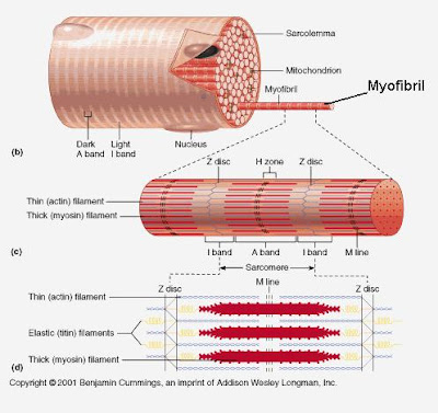
The myofibrils have alternate light and dark bands, which contain protein filaments responsible for the muscle's contractile ability and give the muscle its typical striped appearance under the microscope. The filaments of myofibrils, myofilaments, consist of two types, thick and thin.
Thin filaments consist primarily of the protein actin, coiled with nebulin filaments. Thick filaments consist primarily of the protein myosin, held in place by titin filaments. The protein complex composed of actin and myosin is sometimes referred to as actomyosin.
Tuesday, February 17, 2009
Diastole
Diastole is the period of time when the heart fills with blood after systole. Ventricular diastole is the period during which the ventricles are relaxing, while atrial diastole is the period during which the atria are relaxing. The term diastole originates from Greek and means dilation.
During ventricular diastole, the pressure in the left and right ventricles drops from the peak that it reaches in systole. When the pressure in the left ventricle drops to below the pressure in the left atrium, the mitral valve opens, causing accumulated blood from the atrium to flow into the ventricle.
Myosin
Myosins are a large family of motor proteins found in eukaryotic tissues. They are responsible for actin-based motility. Multiple myosin II molecules generate force in skeletal muscle through a power stroke mechanism fuelled by the energy released from ATP hydrolysis. The power stroke occurs at the release of the products of ATP hydrolysis - ADP and phosphate - when myosin is tightly bound to actin.
Myosin is a protein having a molecular weight of ~ 470,000 daltons. There are about 300 molecules of myosin per thick filament. Each myosin contains two heads that are the site of the myosin ATPase, an enzyme that hydrolyzes ATP required for actin and myosin cross bridge formation. These heads interact with a binding site on actin.
Myosin is a protein having a molecular weight of ~ 470,000 daltons. There are about 300 molecules of myosin per thick filament. Each myosin contains two heads that are the site of the myosin ATPase, an enzyme that hydrolyzes ATP required for actin and myosin cross bridge formation. These heads interact with a binding site on actin.
Systole
Systole is the contraction of heart chambers, driving blood out of the chambers. The chamber most often discussed is the left ventricle. However, all four chambers of the heart undergo systole and diastole in a timed fashion so that blood is propelled forward through the cardiovascular system.
The systolic pressure is specifically the maximum arterial pressure during contraction of the left ventricle of the heart. In a blood pressure reading, the systolic pressure is typically the first number recorded. For example, with a blood pressure of 120/80 ("120 over 80"), the systolic pressure is 120. By "120" is meant 120 mm Hg (millimeters of mercury).
The systolic pressure is specifically the maximum arterial pressure during contraction of the left ventricle of the heart. In a blood pressure reading, the systolic pressure is typically the first number recorded. For example, with a blood pressure of 120/80 ("120 over 80"), the systolic pressure is 120. By "120" is meant 120 mm Hg (millimeters of mercury).
Monday, February 16, 2009
Myocyte
A myocyte is the type of cell found in muscles. From myo-, muscle + -cyte, cell = muscle cell. They arise from myoblasts. Each myocyte contains myofibrils, which are long chains of sarcomeres, the contractile units of the cell. Kindlin-2 plays a role in elongation.
The myocyte is a specialized cardiac muscle cell that is approximately 25 µ in diameter and about 100 µ in length. The myocyte is composed of bundles of myofibrils that contain myofilaments. The myofibrils have distinct, repeating microanatomical units, termed sarcomeres, which represent the basic contractile units of the myocyte. The sarcomere is defined as the region of myofilament structures between two Z-lines.

The myocyte is a specialized cardiac muscle cell that is approximately 25 µ in diameter and about 100 µ in length. The myocyte is composed of bundles of myofibrils that contain myofilaments. The myofibrils have distinct, repeating microanatomical units, termed sarcomeres, which represent the basic contractile units of the myocyte. The sarcomere is defined as the region of myofilament structures between two Z-lines.
Chemical and physical interactions between the actin and myosin cause the sarcomere length to shorten, and therefore the myocyte to contract during the process of excitation-contraction coupling. The interactions between actin and myosin serve as the basis for the sliding filament theory of muscle contraction.
Cardiac Cycle
The cardiac cycle is the sequence of events that occurs when the heart contracts to pump blood throughout the body, specially from the beginning of one heart beat to the next. There are two phases of the cardiac cycle. In the diastole phase, the heart ventricles are relaxed and the heart fills with blood. In the systole phase, the ventricles contract and pump blood into the arteries.
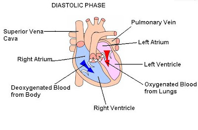

The cardiac cycle begins with a period of fast ventricular filling. The right atrium fills with deoxygenated blood from the superior vena cava, the inferior vena cava, and the coronary venous return. At the same time, the pulmonary veins return oxygenated blood from the lungs to the left atrium. During the early diastolic phase of the cardiac cycle, both ventricles relax and fill from their respective atrial sources. The atrio-ventricular valves (the tricuspid valve and the mitral valve) open and allow blood to flow from the atria into the ventricles.
During the systolic phase, the ventricles contracts to pump the blood out into the arteries. The right ventricle pumps the deoxygenated blood (carbondioxide-containing blood) out into the pulmonary artery, which takes the blood to the lungs to release the carbondioxide and pick up oxygen. The left ventricle pumps the oxygenated blood out into the aortic artery, which distributes the oxygen-rich blood throughout the body.
Sunday, February 15, 2009
Pulmonary Valve
The pulmonary valve, is a three-cusp valve which lies between the right ventricle and the pulmonary artery of the heart. Similar to the aortic valve, the pulmonary valve opens in ventricular systole, when the pressure in the right ventricle rises above the pressure in the pulmonary artery.

The pulmonary valve prevents the back flow of blood as it is pumped from the right ventricle to the pulmonary artery.
Saturday, February 14, 2009
Papillary Muscle
The papillary muscles are small muscles which anchor heart valves. These muscles contract to tighten the chordae tendineae, which in turn prevent inversion. This occurs in response to pressure gradients. Instead the papillary muscles brace the valves against the high pressure, preventing regurgitation of ventricular blood back into the atrial cavities.

The papillary muscles are projections of ventricular muscle to which the tendinous cords, or chordae tendinae, of the atrioventricular valves are attached. Contractions of the papillary muscles assist the tendinous cords in preventing the valves from being thrust out into the atrial cavity during ventricular systole.
Chordae Tendineae
The chordae tendineae, also called heart strings, are cord-like tendons that connect the papillary muscles to the tricuspid valve and the mitral valve in the heart. The chordae tendineae prevents the valves flaps from being everted (turned inside out) into the right atrium when the right ventricle of the heart contracts to pump the blood into the aorta, thus avoiding a backflow of blood into the right atrium.

In the same way, these cord-like tendons hold in position other flaps, such as the bicuspid or mitral valve. Chordae tendineae are approximately 80% collagen, while the remaining 20% is made up of elastin and endothelial cells.
Friday, February 13, 2009
Aortic Valve
The aortic valve is one of the four valves of the heart. It lies between the left ventricle and the aorta. It works like a one-way gate. When the heart pumps during ventricular systole, the aortic valve opens to let oxygen-rich blood flow from the left ventricle into a large blood vessel called the aorta. Blood then flows through the aorta to the rest of the body.

There are two processes that can affect the aortic valve - aortic stenosis in which the valve fails to open fully, thereby obstructing blood flow out from the heart, and aortic insufficiency, also called aortic regurgitation, in which the aortic valve is incompetent and blood flows passively back to the heart in the wrong direction.
Mitral Valve
The mitral valve is a two-flap tissue valve in the heart that lies between the left atrium and the left ventricle. The mitral valve and the tricuspid valve are known collectively as the atrioventricular valves because they lie between the atria and the ventricles of the heart and control the flow of blood.
The mitral valve opens to allow the oxigen-rich blood to flow from the left atrium into the left ventricle, not letting it flow back into the atrium as the heart contracts (systole) to pump this blood to the rest of the body through the aorta.
The mitral valve has two cusp-like leaflets, the anteromedial leaflet and the posterolateral leaflet, which guard the opening. The opening is surrounded by a fibrous ring known as the mitral valve annulus. The orientation of the two leaflets resemble a bishop's miter, whence the valve receives its name.
During left ventricular diastole, after the pressure drops in the left ventricle due to relaxation of the ventricular myocardium, the mitral valve opens, and blood travels from the left atrium to the left ventricle. About 70-80% of the blood that travels across the mitral valve occurs during the early filling phase of the left ventricle. These valve leaflets are prevented from prolapsing (collapsing) into the left atrium by the action of tendons attached to the posterior surface of the valve, chordae tendineae.

Thursday, February 12, 2009
Tricuspid Valve
The tricuspid valve is on the right side of the heart, between the right atrium and the right ventricle. The normal tricuspid valve usually has three leaflets and three papillary muscles. They are connected to the papillary muscles by the chordae tendineae, which lie in the right ventricle. Tricuspid valves may also occur with two or four leaflets, and the number may change during life.

The tricuspid valve prevents the back flow of blood as it is pumped from the right atrium to the right ventricle.
The tricuspid valve can be affected by rheumatic fever, which can cause tricuspid stenosis or tricuspid insufficiency. Some patients are born with congenital abnormalities of the tricuspid valve. Congenital apical displacement of the tricuspid valve is called Ebstein's anomaly and typically causes significant tricuspid regurgitation.
Heart Valves
The heart valves regulate the flow of blood through the heart four chambers. They maintain the unidirectional flow of blood in the heart by opening and closing depending on the difference in pressure on each side.

The heart has four valves: 1) the tricuspid valve is between the right atrium and right ventricle; 2) the pulmonary valve is between the right ventricle and the pulmonary artery; 3) the mitral valve is between the left atrium and left ventricle; 4) the aortic valve is between the left ventricle and the aorta.
Each valve has a set of flaps, also called cusps. When working properly, the heart valves open and close fully. Heart valves don't always work as they should. A person can be born with an abnormal heart valve, a type of congenital heart defect.
Endocardium
The endocardium is the innermost layer of tissue that lines the chambers of the heart and heart valves. The endocardium is thicker in the atria than in the ventricles. Its cells, embryologically and biologically, are similar to the endothelial cells that line blood vessels.

The endocardium consists of a layer of endothelial cells and an underlying layer of connective tissue.
Wednesday, February 11, 2009
Myocardium
The myocardium is the middle muscular layer of the heart wall. It contracts to pump blood out of the heart, then relaxes as the heart refills with returning blood.

The myocardium consists of involuntary striated muscle. Theses muscle cells are known as cardiac myocytes.
Pericardium
The pericardium is a double-walled sac of fibrous tissue that contains the heart and the roots of the great vessels. The pericardium's outer coat is tough and thickened, loosely surrounds the heart, and is attached to the central part of the diaphragm and the back of the breastbone. Its inner coat (the visceral pericardium or epicardium) is double, with one layer closely adherent to the heart and the other lining the inner surface of the outer coat. The intervening space between these layers is filled with pericardial fluid. This small amount of fluid acts as a lubricant to allow normal heart movement within the chest.

The two layers to the pericardial sac are the fibrous pericardium and the serous pericardium. The serous pericardium, in turn, is divided into two layers, the parietal pericardium, which is fused to and inseparable from the fibrous pericardium, and the visceral pericardium, which is part of the epicardium.
Tuesday, February 10, 2009
DNA: Deoxyribonucleic Acid
DNA, or deoxyribonucleic acid, is the hereditary material in humans and almost all other organisms. Nearly every cell in a person’s body has the same DNA. Most DNA is located in the cell nucleus, where it is called nuclear DNA, but a small amount of DNA can also be found in the mitochondria.

Deoxyribonucleic acid (DNA) is a nucleic acid that contains the genetic instructions used in the development and functioning of all known living organisms and some viruses. The main role of DNA molecules is the long-term storage of information. DNA is often compared to a set of blueprints or a recipe, or a code, since it contains the instructions needed to construct other components of cells, such as proteins and RNA molecules. The DNA segments that carry this genetic information are called genes, but other DNA sequences have structural purposes, or are involved in regulating the use of this genetic information.
DNA consists of two long polymers of simple units called nucleotides, with backbones made of sugars and phosphate groups joined by ester bonds. These two strands run in opposite directions to each other and are therefore anti-parallel. Attached to each sugar is one of four types of molecules called bases. It is the sequence of these four bases along the backbone that encodes information. This information is read using the genetic code, which specifies the sequence of the amino acids within proteins. The code is read by copying stretches of DNA into the related nucleic acid RNA, in a process called transcription.
Meiosis
Meiosis is a process of reductional division in which the number of chromosomes per cell is halved. In animals, meiosis always results in the formation of gametes, while in other organisms it can give rise to spores. Before meiosis begins, the DNA in the original cell is replicated. Thus, meiosis starts with homologous chromsomes.

Meiosis is the type of cell division by which germ cells (eggs and sperm) are produced. Meiosis involves a reduction in the amount of genetic material. Meiosis comprises two successive nuclear divisions with only one round of DNA replication.
Meiosis is essential for sexual reproduction and therefore occurs in all eukaryotes that reproduce sexually. A few eukaryotes, notably the Bdelloid rotifers, have lost the ability to carry out meiosis and have acquired the ability to reproduce by parthenogenesis. Meiosis does not occur in archaea or bacteria, which reproduce via asexual processes such as binary fission.
Because meiosis is a "one-way" process, it cannot be said to engage in a cell cycle as mitosis does. However, the preparatory steps that lead up to meiosis are identical in pattern and name to the interphase of the mitotic cell cycle.
Interphase is divided into three phases:
1) Gap 1 (G1) phase: This is a very active period, where the cell synthesizes its vast array of proteins, including the enzymes and structural proteins it will need for growth. In G1 stage each of the chromosomes consists of a single (very long) molecule of DNA. In humans, at this point cells are 46 chromosomes, 2N, identical to somatic cells.
2) Synthesis (S) phase: The genetic material is replicated: each of its chromosomes duplicates, producing 46 chromosomes each made up of two sister chromatids. The cell is still considered diploid because it still contains the same number of centromeres. The identical sister chromatids have not yet condensed into the densely packaged chromosomes visible with the light microscope. This will take place during prophase I in meiosis.
3) Gap 2 (G2) phase: G2 phase is absent in Meiosis.
Interphase is followed by meiosis I and then meiosis II. Meiosis I consists of separating the pairs of homologous chromosome, each made up of two sister chromatids, into two cells. One entire haploid content of chromosomes is contained in each of the resulting daughter cells; the first meiotic division therefore reduces the ploidy of the original cell by a factor of 2.
Meiosis II consists of decoupling each chromosome's sister strands (chromatids), and segregating the individual chromatids into haploid daughter cells. The two cells resulting from meiosis I divide during meiosis II, creating 4 haploid daughter cells. Meiosis I and II are each divided into prophase, metaphase, anaphase, and telophase stages, similar in purpose to their analogous subphases in the mitotic cell cycle.
Monday, February 9, 2009
Cytokinesis
Cytokinesis is the process where the cytoplasm of a single eukaryotic cell is divided to form two daughter cells. It usually initiates during the late stages of mitosis, and sometimes meiosis, splitting a binucleate cell in two, to ensure that chromosome number is maintained from one generation to the next.
Animal cell cytokinesis begins shortly after the onset of sister chromatid separation in the anaphase of mitosis. A contractile ring, made of non-muscle myosin II and actin filaments, assembles in the middle of the cell at the cell cortex, adjacent to the cell membrane. Myosin II uses the free energy released when ATP is hydrolyzed to move along these actin filaments, constricting the cell membrane to form a cleavage furrow. Continued hydrolysis causes this cleavage furrow to move inwards, a striking process that is clearly visible down a light microscope.
Animal cell cytokinesis begins shortly after the onset of sister chromatid separation in the anaphase of mitosis. A contractile ring, made of non-muscle myosin II and actin filaments, assembles in the middle of the cell at the cell cortex, adjacent to the cell membrane. Myosin II uses the free energy released when ATP is hydrolyzed to move along these actin filaments, constricting the cell membrane to form a cleavage furrow. Continued hydrolysis causes this cleavage furrow to move inwards, a striking process that is clearly visible down a light microscope.
Mitosis
Mitosis is the process in which an eukaryotic cell produces two identical daughter cells through nuclear division. Mitosis is followed immediately by cytokinesis, which divides the nuclei, cytoplasm, organelles and cell membrane into two daughter cells containing roughly equal shares of these cellular components.
The process of mitosis is complex and highly regulated. The sequence of events is divided into phases, corresponding to the completion of one set of activities and the start of the next. These stages are prophase, prometaphase, metaphase, anaphase and telophase. During the process of mitosis the pairs of chromosomes condense and attach to fibers that pull the sister chromatids to opposite sides of the cell. The cell then divides in cytokinesis, to produce two identical daughter cells.

Sunday, February 8, 2009
Chromatin
Chromatin is a mass of genetic material composed of DNA and proteins that condense to form chromosomes in eukaryotic cell division. Chromatin is located in the cell's nucleus. The major components of chromatin are DNA and histone proteins, although many other chromosomal proteins have prominent roles too.

The functions of chromatin are to package DNA into a smaller volume to fit in the cell, to strengthen the DNA to allow mitosis and meiosis, and to serve as a mechanism to control expression and DNA replication. Chromatin contains genetic material-instructions to direct cell functions. Changes in chromatin structure are affected by chemical modifications of histone proteins such as methylation and acetylation, and by non-histone, DNA-binding proteins.
Saturday, February 7, 2009
Chromosome
A chromosome is an organized structure of DNA and protein that is found in cells. A chromosome is a single piece of DNA that contains many genes, regulatory elements and other nucleotide sequences.


Chromosomes are found in the cell nucleus. Each one of them contains a single extremely long DNA molecule that is packaged by various proteins into a compact domain. In a human cell nucleus there are 46 chromosomes, or 23 pairs; in every cell except the mature egg and sperm which have a set of only 23 chromosomes (haploid), half the number that is found in ordinary cells.
In eukaryotes, nuclear chromosomes are packaged by proteins into a condensed structure called chromatin. This allows the very long DNA molecules to fit into the cell nucleus. The structure of chromosomes and chromatin varies through the cell cycle. Chromosomes are the essential unit for cellular division and must be replicated, divided, and passed successfully to their daughter cells so as to ensure the genetic diversity and survival of their progeny.
Plasma Membrane
The plasma membrane is the interface between the cellular machinery inside the cell and the fluid outside. It is a selectively permeable lipid bilayer found in all cells The plasma membrane encloses their contents and serves as a semi-porous barrier to the outside environment. The membrane acts as a boundary, holding the cell constituents together and keeping other substances from entering. The plasma membrane is permeable to specific molecules, however, and allows nutrients and other essential elements to enter the cell and waste materials to leave the cell.


The plasma membrane (or cell membrane) surrounds the cytoplasm of a cell and, in animal cells, physically separates the intracellular components from the extracellular environment, thereby serving a function similar to that of skin. The cell membrane also plays a role in anchoring the cytoskeleton to provide shape to the cell, and in attaching to the extracellular matrix to help group cells together in the formation of tissues. The plasma membrane is a barrier which is selectively permeable and able to regulate what enters and exits the cell, thus facilitating the transport of materials needed for survival.
Friday, February 6, 2009
cytosol
The cytosol is the liquid found inside cells. It is the gel-like "soup" within which all the other cell organelles reside and where most of the cellular metabolism occurs. Though mostly water, the cytosol is full of proteins that control cell metabolism including signal transduction pathways, glycolysis, intracellular receptors, and transcription factors. Cytoplasm is a collective term for the cytosol plus the organelles suspended within the cytosol.

The term cytosol is used to refer to the liquid phase of the cytoplasm in an intact cell, this excludes any part of the cytoplasm that is contained within organelles. The cytosol consists mostly of water, dissolved ions, small molecules, and large water-soluble molecules, such as proteins. In contrast to extracellular fluid, cytosol has a high concentration of potassium ions and a low concentration of sodium ions.
Thursday, February 5, 2009
Cytoplasm
The cytoplasm is the parts of a cell that are enclosed within the plasma membrane. It is eighty percent water and usually clear in color. The cytoplasm is a viscous (thick) gel that liquefies when shaken or stirred. Cytoplasm means cell substance. In eukaryotic cells the cytoplasm contains organelles, such as mitochondria, that are filled with liquid kept separate from the rest of the cytoplasm by biological membranes. It is also the site where most cellular activities occur, such as many metabolic pathways, and processes such as cell division.
The part of the cytoplasm that is not held within organelles is called the cytosol. The cytosol is a complex mixture of cytoskeleton filaments, dissolved molecules, and water that fills much of the volume of a cell. The cytosol is a gel, with a network of fibers dispersed through water. Due to this network of pores and high concentrations of dissolved macromolecules, such as proteins, an effect called macromolecular crowding occurs and the cytosol does not act as an ideal solution. This crowding effect alters how the components of the cytosol interact with each other.

The part of the cytoplasm that is not held within organelles is called the cytosol. The cytosol is a complex mixture of cytoskeleton filaments, dissolved molecules, and water that fills much of the volume of a cell. The cytosol is a gel, with a network of fibers dispersed through water. Due to this network of pores and high concentrations of dissolved macromolecules, such as proteins, an effect called macromolecular crowding occurs and the cytosol does not act as an ideal solution. This crowding effect alters how the components of the cytosol interact with each other.

Mitochondrion
A mitochondrion is a membrane-enclosed organelle found in most eukaryotic cells. It is sometimes described as the "cellular power plant" because it generates most of the cell's supply of adenosine triphosphate (ATP), used as a source of chemical energy.

Mitochondria provide the energy a cell needs to move, divide, and produce secretory products. They are the power centers of the cell. They are about the size of bacteria but may have different shapes depending on the cell type. Mitochondria have been implicated in several human diseases, including mitochondrial disorders and cardiac dysfunction, and may play a role in the aging process.
The number of mitochondria in a cell varies widely by organism and tissue type. Many cells have only a single mitochondrion, whereas others can contain several thousand mitochondria.
The mitochondrion is composed of compartments that carry out specialized functions. These compartments or regions include the outer membrane, the intermembrane space, the inner membrane, and the cristae and matrix. Mitochondrial proteins vary depending on the tissues and species. In human, 615 distinct types of proteins were identified from cardiac mitochondria.
Wednesday, February 4, 2009
Ribosome
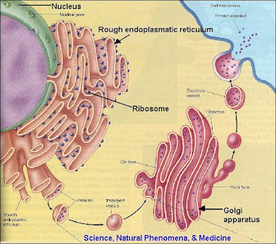
Ribosomes are the protein-synthesizing organelles of the cell. Ribosomes are about 20nm in diameter and are composed of 65% ribosomal RNA and 35% ribosomal proteins, known as a Ribonucleoprotein or RNP. Free ribosomes are suspended in the cytosol, which is the semi-fluid portion of the cytoplasm; others are bound to the rough endoplasmic reticulum, giving it the appearance of roughness and thus its name.
The ribosome functions in the expression of the genetic code from nucleic acid into protein, in a process called translation. Ribosomes do this by catalyzing the assembly of individual amino acids into polypeptide chains; this involves binding a messenger RNA and then using this as a template to join together the correct sequence of amino acids.
Ribosomes were first observed in the mid-1950s by Romanian cell biologist George Palade using an electron microscope as dense particles or granules for which he would win the Nobel Prize. The term "ribosome" was proposed by scientist Richard B. Roberts in 1958.
Tuesday, February 3, 2009
Endoplasmatic Reticulum
The endoplasmatic reticulum is a cell organelle which forms an interconnected network of tubules, vesicles, and cisternae held together by the cytoskeleton within cytoplasm. They play an important role in the synthesis of lipids and proteins to be used in the cell membrane or to be secreted from the cell.
The endoplasmatic reticulum also fulfills other specialized functions to such as sequestration of calcium, and production and storage of glycogen, steroids, and other macromolecules. There are two types of endoplasmatic reticulum: smooth and rough.
Smooth endoplasmatic reticulum is the site of synthesis and digestion of fatty acids and phospholipids. In the liver it is used to modify dangerous chemicals. Rough ER is the site of manufacture of secretory proteins as well as proteins destined to be inserted in the cell membrane. It is rough because of the vast numbers of ribosomes which stud its surface. As these ribosomes build an amino acid chain, it is injected through the endoplasmatic reticulum into the vesicle.
The lacey membranes of the endoplasmic reticulum were first seen by Keith R. Porter, Albert Claude, and Ernest F. Fullam in 1945.

Subscribe to:
Posts (Atom)