Sunday, May 31, 2009

Occipital Lobe

The occipital lobe is the most posterior portion of the cerebral cortex of the human brain, at the back of the head. It is the smallest of the four lobes of each cerebral hemisphere and is separated from the parietal lobe by calcarine fissure. The occipital lobe is defined as the part of the cerebral cortex that lies underneath the occipital bone.

The occipital lobe is the visual center of the brain as it contains the primary visual cortex. Each visual cortex receives sensory information from the outside half of the retina on the same side of the head and from the inside half of the retina on the other side of the head. The cuneus, also called the Brodmann's area 17, receives visual information from the contralateral superior retina representing the inferior visual field. The lingula receives information from the contralateral inferior retina representing the superior visual field. The retinal inputs pass through a "way station" in the lateral geniculate nucleus of the thalamus before projecting to the cortex.


Saturday, May 30, 2009

Prefrontal Cortex

The prefrontal cortex is the anterior portion of the frontal lobes of the cerebrum. It lies in front of the motor and premotor areas. The prefrontal cortex is thought to be involved in planning complex cognitive behavior and in the expression of personality and appropriate social behavior. The prefrontal cortex can be divided into three basic areas: the orbitofrontal and ventromedial areas; the dorsolateral prefrontal cortex; the anterior and ventral cingulate cortex.

The prefrontal cortex has been shown to participate in the association of events separated by time and is responsible for the executive functions, which include mediating conflicting thoughts, making choices, and governing social control. When the pathways between the prefrontal cortex and the rest of the brain are damaged due to head injury, massive personality changes can result.

Many diseases, such as schizophrenia, bipolar disorder and ADHD, have been related to dysfunction of the prefrontal cortex, and thus this area of the brain offers the potential for new treatments of these diseases. Markers of inhibitory neurotransmission are altered in the prefrontal cortex (PFC) of subjects with schizophrenia, and several lines of evidence suggest that these alterations may be most prominent in the subset of GABA-containing neurons that express the calcium-binding protein, parvalbumin (PV).

The prefrontal cortex responds mostly to stimuli signaling the need for movement; nevertheless it is also responsible for many other specialized functions. It gets information from all sensory systems and can integrate a large amount of information. It has been shown that the prefrontal cortex is responsible for working memory. Working memory is defined as the information that is currently available in memory for working on a problem.

Friday, May 29, 2009

Frontal Lobe

The frontal lobe is the anterior portion of the cerebral cortex of humans. It is largest lobe of each cerebral hemisphere and is separated from the parietal lobe by the central sulcus of Rolando and from the temporal lobe by the lateral sulcus. The frontal lobe is responsible for the control of skilled motor activity, including speech.

In the frontal lobe, there are four important gyri. Roughly parallel and anterior to the central sulcus is the Precentral Gyrus, which is also called the motor strip. The precentral gyrus represents the primary motor cortex. This motor cortical area contains motor neurons whose axons extend to the spinal cord and brain stem and synapse on motor neurons in the spinal cord. Besides the roughly vertical Precentral Sulcus (anterior to the precentral gyrus), there are two additional sulci that are sort of diagonal. These sulci provide boundaries for important frontal gyri: the Superior Frontal Gyrus, the Middle Frontal Gyrus, and the Inferior Frontal Gyrus. The most anterior region of the frontal lobe is called the prefrontal cortex and includes all three of these gyri.

The frontal lobe integrates and coordinates all the other regions of the cerebral cortex and is the emotional control center and home to our personality. Injuries sustained by the frontal lobe can cause a wide variety of symptoms. The frontal lobe is involved in motor function, problem solving, spontaneity, memory, language, initiation, judgement, impulse control, and social and sexual behavior. The frontal lobes are extremely vulnerable to injury due to their location at the front of the cranium, proximity to the sphenoid wing and their large size. MRI studies have shown that the frontal area is the most common region of injury following mild to moderate traumatic brain injury.

There are asymmetrical differences between the two frontal lobes. The left frontal lobe is involved in controlling language related movement, while the right frontal lobe plays a role in non-verbal abilities. The executive functions of the frontal lobes include the capacity to recognize future consequences resulting from current actions, to suppress unacceptable social responses, choose between good and bad actions, and determine similarities and differences between things or events. In other words, it is involved in higher mental functions.

The frontal lobe contains most of the dopamine-sensitive neurons in the cerebral cortex. The dopamine system is associated with reward, attention, long-term memory, planning, and drive. Dopamine tends to limit and select sensory information arriving from the thalamus to the fore-brain. The frontal lobe reaches full maturity around age 25, marking the cognitive maturity associated with adulthood. It has been found increased myelin in the frontal lobe white matter of young adults compared to that of teens. A typical onset of schizophrenia in early adult years correlates with poorly myelinated and thus inefficient connections between nerve cells in the fore-brain. The frontal lobe of the brain acts like a cerebral projecting lens as mind that governs brain. When the lens-like frontal function fails, as in schizophrenia, the mind disintegrates.


Thursday, May 28, 2009

Apical Dendrite

An apical dendrite is a dendrite which projects from the apex of a pyramidal neuron. The apical dendrite belongs to one of two primary categories of dendrites, and it distinguishes the pyramidal neuron from the spiny stellate cell in the cortices. Pyramidal cells are found in the prefrontal cortex, the hippocampus, the entorhinal cortex, and the olfactory cortex. Dendrite arbors formed by apical dendrites are the means by which synaptic inputs into a cell are integrated.

The apical dendrites in these regions contribute significantly to memory, learning, and sensory associations by modulating the excitatory and inhibitory signals received by the pyramidal neurons. Apical dendrites can be divided into two categories: distal and proximal. The longer distal apical dendrites project from the pyramidal neuron body opposite from the axon. Distal apical dendrites form non-local synapses. Shorter proximal apical dendrites project radially to local pyramidal cells and interneurons. Pyramidal neurons segregate their inputs using proximal and apical dendrites.

Wednesday, May 27, 2009

Pyramidal Neuron

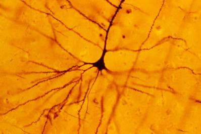
The pyramidal neuron is a type of neuron found in areas of the brain including cerebral cortex, the hippocampus, and in the amygdala. Pyramidal neurons are the main excitation units of the prefrontal cortex and the corticospinal tract in human beings. Pyramidal neurons were first discovered and studied by Santiago Ramón y Cajal. Since then, studies on pyramidal neurons have focused on topics ranging from neuroplasticity to cognition.

One of the key structural features of the pyramidal neuron is the triangular shaped neuron body, which is called soma; hence the name of the neuron. Other important structural features of the pyramidal cell are a single axon, a large apical dendrite, a basal dendrite, and the presence of dendritic spines.

Thousands of pyramidal neurons in sensory, motor, association and executive cortex reveal marked differences in the numbers of putative excitatory inputs received by these cells. Pyramidal nerve cells in prefrontal cortex have, on average, up to 23 times more dendritic spines than those in the primary visual area. It has been proposed that without these specializations in the structure of pyramidal cells, and the circuits they form, human cognitive processing would not have evolved to its present state. Scientific data from both New World and Old World monkeys show varying degrees of complexity in the pyramidal neuron phenotype in their prefrontal cortices, suggesting that cortical circuitry and, thus, cognitive styles are evolving independently in different species.

Pyramidal neurons have been classified into different subclasses based upon their firing responses to 400-1000 millisecond current pulses. These classification are RSad, RSna, and IB neurons. RSad pyramidal neurons, or adapting regular spiking neurons, fire with individual action potentials (APs), which are followed by a hyperpolarizing afterpotential. The afterpotential increases in duration which creates spike frequency adaptation in the neuron. RSna pyramidal neurons, or non-adapting regular spiking neurons, fire a train of action potentials after a pulse. These neurons fail to show any signs of adaptation.

Tuesday, May 26, 2009

Neocortex

The neocortex is the part of the cerebrum which forms the outer layer of the cerebral hemispheres. The neocortex consists of six layers neurons, labelled I to VI, with VI being the innermost and I being the outermost. It is the center of higher mental functions, such as sensory perception, generation of motor commands, spatial reasoning, conscious thought, and language. The neocortex contains about 100 billion cells, each with 1,000 to 10,000 synapses. "Neocortex" means in Latin "new bark."

The neocortex is composed of the grey matter, which is made up of the neuronal cell bodies and unmyelinated axons. It surrounds the deeper white matter (myelinated axons) in the cerebrum. The neocortex is smooth in rodents and other small mammals, but it has deep grooves (sulci) and wrinkles (gyri) in primates and specially human beings. These deep folds increase the surface area of the neocortex considerably without taking up too much more volume.

The neocortex is formed by two primary types of neurons, excitatory pyramidal neurons, which make up about 80% of neocortex, and inhibitory interneurons (~20%). The structure of the neocortex is relatively uniform. It consists of six horizontal layers segregated principally by cell type and neuronal connections. The neocortex is divided into frontal, parietal, temporal, and occipital lobes, which perform different functions. The occipital lobe contains the primary visual cortex, and the temporal lobe contains the primary auditory cortex. Further subdivisions or areas of neocortex are responsible for more specific cognitive processes. In humans, the frontal lobe contains areas devoted to abilities that are unique to our species, such as complex language processing localized to the ventrolateral prefrontal cortex (Broca's area) and social and emotional processing localized to the orbitofrontal cortex.

The neocortex is the newest part of the cerebral cortex to evolve (hence the name "neo"); the other parts of the cerebral cortex are the paleocortex and archicortex, collectively known as the allocortex. The cellular organization of the allocortex is different from the six-layer structure mentioned above. In humans, 90% of the cerebral cortex is neocortex.

Monday, May 25, 2009

Ventricular System

The ventricular system is a series of interconnected, fluid-filled cavities in the brain. These cavities are continuous with the central canal of the spinal cord. The ventricular system consists of four ventricles: two lateral ventricles, right and left; the third ventricle; and the fourth ventricle. There are a few little holes, which are called foramen, in the brain leading from these ventricles.

The ventricular spaces in the various subdivisions of the brain reflects the fact that the ventricles are the adult derivatives of the open space of the embryonic neural tube. The two lateral ventricles, situated within the cerebrum, are relatively large and C-shaped, and roughly wraps around the dorsal aspects of the basal ganglia. In the lateral ventricles of the embryo the successive generation of neurons gives rise to the 6-layered structure of the neocortex, constructed from the inside out during development.

The third ventricle forms a narrow midline cavity between the right and left thalamus, communicating with the lateral ventricles through a small opening at the anterior end of the third ventricle. The third ventricle is continuous caudally with the cerebral aqueduct, which runs though the midbrain. At its caudal end, the aqueduct opens into the fourth ventricle, a larger space in the dorsal pons and medulla.


The Ventricular System of the Brain


Sunday, May 24, 2009

Obex

The obex is the point in the human brain at which the fourth ventricle narrows to become the central canal of the spinal cord. It is a small triangular membrane at the caudal end of the roof of the fourth ventricle of the brain. The decussating of sensory fibers happens at this point. Word "obex" derives from the Latin and means "barrier."

Sulcus Limitans

The sulcus limitans is a slight depressions in the fourth ventricle separating the basal and alar plates. It forms the lateral boundary of the medial eminence. In the superior part of the rhomboid fossa, the sulcus limitans corresponds with the lateral limit of the fossa and presents a bluish-gray area, the locus ceruleus.


Saturday, May 23, 2009

Facial Colliculus

The facial colliculus is a raised area which lies on the dorsal pons. It consists of motor fibers of the facial nerve as they loop over the abducens nucleus. In other words, the facial colliculus is a bump in the floor of the fourth ventricle caused by the motor fibers of the cranial nerves VII looping over the abducens nucleus.


Friday, May 22, 2009

Lateral Ventricles

The lateral ventricles are cavities that are part of the ventricular system of the brain. They are located in the lower parts of the cerebral hemispheres. They are the largest of the ventricles and each consists of a central part, with anterior, posterior and inferior horns. The lateral ventricles are connected to the third ventricle through the interventricular foramina of Monro.


Thursday, May 21, 2009

Fourth Ventricle

The fourth ventricle is one of the cavities of the ventricular system which lies at base of the cerebrum. It is filled with cerebrospinal fluid and extends from the aqueduct of Sylvius to the obex. In cross-section of the human brain, the fourth ventricle is diamond-shaped.

The roof of the fourth ventricle is formed by the cerebellum (superior and inferior medullary vela), the floor by the rhomboid fossa, and the side "walls" formed by the cerebellar peduncles. Among the prominent features of the floor of the fourth ventricle are the: facial colliculus, which is formed by the internal part of the facial nerve as it loops around the abducens nucleus in the lower pons; the sulcus limitans, which represents the border between the alar plate and the basal plate of the developing neural tube; and the obex, which represents the caudal tip of the fourth ventricle and is also a marker for the level of the foramen magnum of the skull and therefore is a marker for the imaginary dividing line between the medulla and spinal cord.

Hypothalamus

The hypothalamus is the part of the brain which lies below the thalamus, in the sella turcica of the cranium, and forms the portion of the ventral region of the diencephalon. The main function of the hypothalamus is to regulate body temperature, blood pressure, heartbeat, certain metabolic processes, and other activities of the Autonomic Nervous System. It also regulates a variety of hormonal secretion by action on the pituitary gland, and controls the contraction and relaxation of the blood vessels walls.

The hypothalamus is a part of the limbic system, which influences important aspects of our behavior and even our very survival, regulating such functions as emotion, sexual drives, nutritional appetites, rhythms, and sleep cycles. The frontal lobes of the cerebral cortex connects to the hypothalamus. Disturbances in these pathways are thought to result in abnormal affective behavior.

Wednesday, May 20, 2009

Third Ventricle

The third ventricle, or ventriculus tertius, is a narrow cavity located between the two cerebral hemispheres. It is one of four connected fluid-filled cavities which comprise the ventricular system within the cerebrum of human beings. The third ventricle is in the midline, between the left and right lateral ventricles and is filled with cerebrospinal fluid.

The third ventricle is bounded by the thalamus and hypothalamus on both the left and right sides. The third ventricle provides a pathway for cerebrospinal fluid.

Tuesday, May 19, 2009

Thalamus

The thalamus is an ovoid mass of gray matter located in the posterior part of the forebrain. It relays sensory impulses to the cerebral cortex. The thalamus processes and relays sensory information selectively to different parts of the cerebral cortex, as one thalamic point may reach one or several regions in the cortex.

The thalamus is made up of neurons bodies and is situated lateral to the third ventricle that serves as the principal relay and integration station for the sensory systems in the body. Together with the hypothalamus, the thalamus establishes levels of sleep and wakefulness. It is also vital to the neural feedback system controlling brain wave rhythms.


Hippocampus

The hippocampus is an elevation of the floor of each lateral ventricle of the cerebrum which consists mainly of gray matter and is covered by a layer of white matter. It is part of the lymbic system and has a central role in memory processes.

The hippocampus can be divided into two parts: hippocampus major, or horn of Ammon, and hippocampus minor. The hippocampus is closely related to the cerebral cortex, and is located in the medial temporal lobe, underneath the cortical surface. It is shaped like a curved tube, which in humans is convoluted in a way that reminded early anatomists of a seahorse ("hippocampus" means "seahorse" in Greek).

Neuroscientists agree that the hippocampus plays an important role in the formation of new memories about experienced events. Some researchers view the hippocampus as part of a larger medial temporal lobe memory system responsible for long term memory. Severe damage to the hippocampus results in profound difficulties in forming new memories (anterograde amnesia), and often also affects memories formed prior to the damage (retrograde amnesia). Although the retrograde effect normally extends some years prior to the brain damage, in some cases older memories remain. This sparing of older memories leads to the idea that consolidation over time involves the transfer of memories out of the hippocampus to other parts of the brain. Damage to the hippocampus does not affect some types of memory, such as the ability to learn new motor or cognitive skills.


Monday, May 18, 2009

Globus Pallidus

The globus pallidus is a major constituent of the basal ganglia core, which is a subcortical structure of the brain. The globus pallidus is divided into two segments by the medial medullary lamina: internal globus pallidus, and external globus pallidus, which are surrounded everywhere by myelinic walls. Both receive input from the caudate nucleus and putamen, and both are in communication with the subthalamic nucleus.

The globus pallidus, like the rest of the basal ganglia, is grey matter, but a light grey, made up neurons bodies and is surrounded by white matter. The globus pallidus is traversed by the numerous myelinated axons of the striato-pallidonigral bundle that give it the pale appearance from which it is named. The globus pallidus is one of three nuclei that make up the basal ganglia and is involved in the regulation of voluntary movements at a subconscious level.

Substantia Nigra

The substantia nigra is a brain structure which is part of the basal ganglia. It is made up of neurons and plays an important role in movement and addiction. The substantia nigra consists of two parts with very different connections and functions; the pars compacta and pars reticulata. The pars compacta serves mainly as an input to the basal ganglia circuit and supplies the striatum with dopamine. The pars reticulata, on the other hand, serves mainly as an output, conveying signals from the basal ganglia to numerous other brain structures.

The destruction of the substantia nigra is associated with Parkinson's disease. Parkinson's disease's is a neurodegenerative disease caused by the death of dopaminergic neurons in the pars compacta of the substantia nigra. The cause of death of dopaminergic neurons in the pars compacta is unknown.

Sunday, May 17, 2009

Caudate Nucleus

The caudate nucleus is a nucleus located within the basal ganglia of the human brain. The caudate nucleus is a collection of neurons bodies that connects to many parts of the cerebrum. Together with the putamen, it comprises the striatum. It is involved with control of voluntary movement, and the deterioration of its connections results in the inability to control movements and emotions.

The caudate nuclei are located near the center of the brain, sitting astride the thalamus. There is a caudate nucleus within each hemisphere of the brain. Individually, they resemble a C-shape structure. The head and body of the caudate nucleus form part of the floor of the anterior horn of the lateral ventricle.

The caudate organizes and filters information that is sent to the frontal lobe, particularly information from the limbic system. Caudate malfunction can affect the functioning of the frontal lobes through a lack of information or an improper amount of information.

Saturday, May 16, 2009

Putamen

The putamen is a round structure, which is part of the basal ganglia that is located at the base of the cerebrum. The putamen and caudate nucleus together form the dorsal striatum, which is one of the structures that comprises the basal ganglia. Through various pathways, the putamen is connected to the substantia nigra and globus pallidus.

The main function of the putamen is to regulate movements and influence various types of learning. It employs dopamine to perform its functions. The putamen also plays a role in degenerative neurological disorders, such as Parkinson's disease. The caudate works with the putamen to receive the input from cerebral cortex. They can be considered the "entrance" to the basal ganglia. The nucleus accumbens and medial caudate receive input from frontal cortex and limbic regions. The putamen and caudate are jointly connected with the substantia nigra, but most of their output goes to the globus pallidus.


Basal Ganglia

The basal ganglia are a group of grey nuclei in the brain made up of neurons bodies which send their axons to interconnect with the cerebral cortex, thalamus and brainstem. The basal ganglia are associated with a variety of functions: motor control and emotions.

There are five individual nuclei that make up the human basal ganglia. The rostral basal ganglia is divided into the striatum, which contains the putamen and caudate nucleus, the external segment of the globus pallidus, and the internal segment of the globus pallidus. The caudal basal ganglia comprise the subthalamic nucleus and the substantia nigra. There are 2 sets of basal ganglia in the mammalian brain, mirrored in the left and right hemispheres.

The striatum is the main input zone for other brain areas to connect to the basal ganglia. Via the striatum, the basal ganglia receives input from the cortex, mainly from the motor and prefrontal cortices. The basal ganglia and cerebellum are large collections of nuclei that modify movement on a minute-to-minute basis. Motor cortex sends information to both, and both structures send information right back to cortex via the thalamus. The output of the cerebellum is excitatory, while the basal ganglia are inhibitory. The balance between these two systems allows for smooth, coordinated movement, and a disturbance in either system will show up as movement disorders.


Thursday, May 14, 2009

Cerebrum

The cerebrum together with the diencephalon, constitute the forebrain. It is the most superior region of the vertebrate central nervous system. The cerebrum is alson known as the"telencephalon", which refers to the embryonic structure, from which the mature "cerebrum" develops.

The cerebrum is divided into two symmetric cerebral hemispheres; left and right hemispheres. The cerebrum lies on top of the brainstem and is the largest and most well-developed of the five major divisions of the brain. It is the newest structure in the phylogenetic sense, with mammals having the largest and most well-developed among all species. The cerebrum is composed of the cerebral cortex, basal ganglia, and the olfactory bulb. In human beings, the cerebral cortex is folded into many gyri and sulci, which has allowed the cortex to expand in surface area without taking up much greater volume.

The cerebrum surrounds older parts of the brain. Motor areas of the cerebral cortex as well as limbic and olfactory systems project fibers from the cerebrum to the brainstem and spinal cord. Fibers connect the cognitive and volitive systems of the cerebrum to the thalamus and other regions of the midbrain. The cerebrum's networks of neurons make possible complex behavior such as social interactions, speech and language, learning, memory, and planning.

The cerebrum is also the center of consciousness and volition (will), located in the frontal lobe. Upper motor neurons in the primary motor cortex send their axons to the brainstem and spinal cord to synapse on the lower motor neurons that contract the muscles. Damage to motor areas of cortex can lead to certain types of motor neuron disease such as paralysis.

Language is attributed to certain parts of the cerebral cortex. Motor portions of language are attributed to Broca's area within the frontal lobe; this area is responsible for the dextrous and precise movement of the tongue. The Wernicke's area, at the temporal-parietal lobe junction, is responsible for speech comprehension. The arcuate fasciculus, a large white matter tract, interconnects these two regions. Damage to the Broca's area can result in expressive aphasia, while damage to Wernicke's area results in receptive aphasia.


Wednesday, May 13, 2009

Cerebral Cortex

The cerebral cortex is the highly developed brain structure which plays a key role in consciousness, memory, language, attention, perceptual awareness, and thought. The cerebral cortex is the massive outgrowth of the central nervous system. It is the product of millions of years of evolution.

The cerebral cortex is the highly convoluted external surface of the brain packed in tightly in the cranium. This inordinately creased and folded brain surface arose during evolution as the volume of the cortex increased considerably more rapidly than the cranial volume. The cerebral cortex consists of more than 100 billion neurons. More than two-thirds of the cortical surface is buried in the grooves, called "sulci" and "fissures;" with the latter being deeper than the former. It has a grey color, hence the name "grey matter". Grey matter is formed by neurons and their unmyelinated fibers.

In evolutionary terms, the most recent part of cerebral cortex is called the neocortex, which is made up of six layers of neurons; this is the seat of human intelligence and thoughts. The more ancient part of the cerebral cortex is called the hippocampus, which has three cellular layers, and is divided into subfields.

The cerebral cortex is divided into right and left hemispheres. It encompasses about two-thirds of the brain mass and lies over and around most of the other brain structures. The fissures divide the cerebral cortex into lobes; frontal, temporal, parietal, and occipital. Each one of these lobes have a specific function. For example, there are specific areas involved in vision, hearing, touch, movement, and smell. Other areas are critical for thinking and reasoning.


Melatonin

Melatonin is a hormone secreted by the pineal gland at the base of the brain. It plays an important role in regulating sleep and in maintaining circadian rhythm, the body's natural time clock. The hypothalamus keeps track of the amount of sunlight that is taken in by the eye. The less sunlight, the more melatonin that is released by the pineal gland, thereby enhancing and regulating sleep. Melatonin can also be taken in an over-the-counter supplement mainly sold in health food stores and pharmacies.

Melatonin also exerts a powerful antioxidant activity. The discovery of melatonin as an antioxidant was made in 1993.

Tuesday, May 12, 2009

Gamma Aminobutyric Acid

Gamma aminobutyric acid is an inhibitory neurotransmitter in the central nervous system. It regulates neuronal excitability throughout the nervous system. The Gamma Amino Butiric Acid is directly responsible for the regulation of muscle tone.

Gamma aminobutyric acid is the product of a biochemical decarboxylation reaction of glutamic acid by the vitamin pyridoxal, as well as from decarboxylase. Gamma aminobutyric acid blocks the transmission of a nervous signal from one cell to another in the central nervous system, preventing over-firing of the nerve cells.

Gamma aminobutyric acid is used to treat both epilepsy and hypertension where it is thought to induce tranquility in individuals who have a high activity of manic behavior and acute agitation.

Sunday, May 10, 2009

Dopamine

Dopamine is a metabolic neurotransmitter which is produced in several areas of the brain that include the substantia nigra and the ventral tegmental area. It is the main neurotransmitter in extrapyramidal tracts. High dopamine levels have been linked to aggression.

In the brain, this phenethylamine functions as a neurotransmitter, activating the five types of dopamine receptors — D1, D2, D3, D4 and D5, and their variants. Dopamine is also a neurohormone released by the sympathetic ganglia in the hypothalamus. Its function as a hormone is to inhibit the release of prolactin from the anterior lobe of the pituitary.

In neurons, dopamine is packed into vesicles, which are then released into the synapse in response to a presynaptic action potential. When supplied as a medication, dopamine acts on the sympathetic nervous system and produces effects such as increased heart rate and blood pressure.

Dopamine has many functions in the brain, including important roles in behavior and cognition, motor activity, motivation and reward, inhibition of prolactin production, which is involved in lactation, sleep, mood, attention, and learning.

Saturday, May 9, 2009

Glutamate

Glutamate is a salt of glutamic acid, which functions as an excitatory neurotransmitter. It is the most common excitatory neurotransmitter in the Central Nervous System. Glutamate is stored in vesicles for chemical synapses. Nerve signals activate the release of glutamate from the pre-synaptic cell. In the opposing post-synaptic cell, glutamate receptors bind glutamate. Because of its synaptic plasticity, scientists believe that glutamic acid is involved in cognitive functions like learning and memory in the brain.

Glutamate increases the flow of positive ions by opening ion-channels. Glutamate stimulation is terminated by a membrane transport system that is only used for re-absorbing glutamate & aspartate across the pre-synaptic membrane.


Saltatory Conduction

Saltatory conduction is the means by which a nerve impulse is conducted rapidly through myelinated axons. The nerve signal appears to leap from one node of Ranvier to the next, rather than traveling the entire length of the axon. AS the cytoplasm of the axon is electrically conductive, depolarization at one node of Ranvier is sufficient to elevate the voltage at a neighboring node to the threshold for action potential initiation. Thus in myelinated axons, action potentials do not propagate as waves, but recur at successive nodes and in effect "hop" along the axon, by which process they travel faster than they would otherwise.

Thursday, May 7, 2009

Node of Ranvier

Nodes of Ranvier are the 1-micrometer gaps formed along the length of the myelin-wrapped axons of the nerve cells. The nodes allow the nerve impulses to jump from one node to the next. This is called saltatory conduction. Neurons axons are encased in a sheath of many-layered fatty coating called myelin, which is produced by the Schwann cells. This axonal membrane is uninsulated at nodes of Ranvier and therefore capable of generating electrical activity.

Wednesday, May 6, 2009

Serotonin

Serotonin is a monoamine neurotransmitter synthesized in serotonergic neurons in the central nervous system and enterochromaffin cells in the gastrointestinal tract of animals including humans. As a neurotransmitter, serotonin plays an important role in the modulation of anger, aggression, body temperature, mood, sleep, human sexuality, appetite, and metabolism. Thus, an increase in serotonin levels have a calming effect, relieving depression, insomnia, and irritability. Low serotonin levels are associated with wakefulness and greater sensitivity to pain.

Central Nervous System

The Central Nervous System is the part of the Nervous System which consists of the cerebrum, cerebelum, cerebral peduncles, pons, medulla oblongota, and spinal cord. It is encased within the cranium and the spinal column. The Central Nervous System contains important centers of life (hypothalamus, thalamus, amygdala, pons), playing a central role in the control of most bodily functions, and intelligence and behavior (cerebral cortex).

Tuesday, May 5, 2009

Noradrenaline

Noradrenaline, or norepinephrine, is a neurotransmitter which is produced and released by the neurons of the peripheral sympathetic nervous system. It is responsible for the adrenergic neurotransmission at nerve endings in the heart, muscle, and in many glands, directly increasing the heart rate and blood flow to skeletal muscle and triggering the release of glucose from energy stores.

Noradrenaline is synthesized from dopamine by dopamine ß-hydroxylase and is released from the adrenal medulla into the blood as a hormone. Tyrosine, an amino acid present in body fluids, is taken up into the adrenergic nerve terminal where it is acted upon by an enzyme, tyrosine hydroxylase, to form DOPA; this is converted to dopamine and in turn to noradrenaline. The neurotransmitter is stored in small vesicles, awaiting release.

Noradrenaline is also an important transmitter in many parts of the central nervous system, where it is involved in arousal, blood pressure regulation, and mood. The noradrenergic neurons in the brain form a neurotransmitter system, which exerts effects on large areas of the brain. The effects are alertness and arousal, and influences on the reward system.

Monday, May 4, 2009

Synapse

Synapse is the gap between the axon terminals of one neuron and the dendrite, or cell body, of another. It is at this cleft point that the electrochemical signals of one neuron, coming along its axon length, jumps the gap onto the other neuron's dendrite. The springboard for this signal jump is a chemical substance called neurotransmitter, which makes possible this synapse phenomenon. Synapse also exists between a neuron and a non-neuronal cell, such as those in muscles or glands.

The word synapse was first used at the end of the nineteenth century by the British neurophysiologist Charles Sherrington, who argued, on the basis of his own observations of reflex responses and the studies of the great Spanish anatomist, Ramón y Cajal, that a special form of transmission takes place at the contact between one nerve cell and the next.
Although most synapses connect axons to dendrites, there are also other types of connections, including axon-to-cell-body, axon-to-axon, and dendrite-to-dendrite. Synapses are generally too small to be recognizable using a light microscope. They can only be visualized clearly using an electron microscope.

Synapses send information-containing impulses directionally from a presynaptic neuron to a postsynaptic one. The presynaptic terminal, or synaptic button, is a specialized area within the axon of the presynaptic nerve cell which contains neurotransmitters enclosed in small membrane-bound spheres called synaptic vesicles. These synaptic vesicles are docked at the presynaptic plasma membrane at regions called active zones.

The synaptic process starts with a wave of electrochemical excitation called an action potential which travels along the membrane of the presynaptic cell, until it reaches the synaptic gap. The electrical depolarization of the membrane at the synapse causes channels to open that are permeable to calcium ions.

Calcium ions flow through the presynaptic membrane, rapidly increasing the calcium concentration in the interior. The high calcium concentration activates a set of calcium-sensitive proteins attached to vesicles that contain a neurotransmitter chemical. These proteins change shape, causing the membranes of some docked vesicles to fuse with the membrane of the presynaptic nerve cell, opening the vesicles and dumping their neurotransmitters into the synaptic cleft, the narrow space between the membranes of the pre- and post-synaptic cells.

The neurotransmitter diffuses within the cleft. Some of it escapes, but some of it binds to chemical receptor molecules located on the membrane of the postsynaptic neuron. The binding of neurotransmitter causes the receptor molecule to be activated in some way. Due to thermal shaking, neurotransmitter molecules eventually break loose from the receptors and drift away. The neurotransmitter is either reabsorbed by the presynaptic nerve cell, and then repackaged for future release, or else it is broken down metabolically.


Sunday, May 3, 2009

Neuron

A neuron is an excitable cell which is the functional unit of the nervous system. Also known as nerve cell, it processes and transmits information by electrochemical signalling from neuron to the next via synapse. Neurons are the core components of the brain, the vertebrate spinal cord, and the peripheral nerves.

The neuron consists of a cell body, which is called soma, and one or more long processes: a single axon and dendrites. The soma contains the nucleus and usual cytoplasmic organelles with an exceptionally large amount of rough endoplasmic reticulum, called Nissl substance in the neuron. The longest cell process is the axon, which is capable of transmitting propagated nerve impulses.

Neurons are highly specialized for the processing and transmission of electrochemical impulses generated by neurotransmitters which are secreted by the soma and released at the terminal ends of the axons during synaptic jumps.

The neuron's role as the primary functional unit of the nervous system was first discovered by the Spanish anatomist Santiago Ramón y Cajal in the early 20th century. Cajal wrote that neurons were discrete cells that communicated with each other via specialized junctions (synaptic gaps) between cells.

When a neuron has no dendrite, it is called unipolar neuron; when it has one dendrite, it is a bipolar neuron; if it has more than one dendrite, it is a multipolar neuron. In every neurons only the axon propagates nerve impulses.


Acetylcholine

Acetylcholine is a neurotransmitter in both the central nervous system and peripheral nervous system of vertebrates. It is a white crystalline derivative of choline, C7H17NO3, that is released at the end of axons and is involved in the transmission of nerve impulses in the body. In the cerebral cortex, it is important in memory and learning processes.

Acetylcholine carries nerve impulses across the synaptic gap, from one neuron to another. It functions as a chemical messager whose message is read via receptors in the neurons and muscle tissues. It is active at many nerve synapses and at the motor end plate of vertebrate voluntary muscles.

In the peripheral nervous system, acetylcholine activates muscles, and is a major neurotransmitter in the autonomic nervous system. In the central nervous system, acetylcholine and the associated neurons form a neurotransmitter system, the cholinergic system, which tends to cause excitatory actions.

Saturday, May 2, 2009

Neurotransmitters

Neurotransmitters are chemical substances produced by the cell body of the neurons. Then they are transported to the terminal ends of the neuron, axon teledendria buttons where they are stored. Neurotransmitters allow the movement of information from one neuron to the next, across the synaptic gaps between axons and dendrite. This phisiological phenomenom is called synapse. The neurotransmitters diffuse across the gap to bind with receptors on the postsynaptic cell membrane and cause electrical changes in that neuron.

The most important neurotransmitters are: acetylcholine, dopamine, serotonin, noradrenaline, endorphin, glutamate, and gamma aminobutyric acid. Neurotransmitter are broken down once it reaches the post-synaptic cell to avoid further excitatory or inhibitory signal transduction. Acetylcholine, which is an excitatory neurotransmitter, is broken down by acetylcholinesterase. Choline is taken up and recycled by the pre-synaptic neuron to synthesize more acetylcholine. Other neurotransmitters, such as dopamine, diffuse away from their targeted synaptic junctions and are eliminated from the body via the kidneys, or destroyed in the liver.