Friday, March 27, 2009

Astigmatism

Astigmatism is an optical defect in which the sight is blurred because of the incapacity of the optical system of the eye to focus the image of an object sharply to a point on the retina.

Astigmatism occurs when the cornea, or the eye lens, has an irregular curvature, and it is one of a group of eye conditions known as refractive errors.

There are two kinds of astigmatism; regular and irregular. Regular astigmatism is an alteration in the normal shape of the cornea, or lens. Irregular astigmatism is is caused by a corneal scar or scattering in the crystalline lens.

Astigmatism can be corrected with eyeglasses, contact lenses, or refractive surgery.

Thursday, March 26, 2009

Ciliary Disk

The ciliary disk, or orbiculus ciliaris, is a circular tract in the eye that extends from the ora serrata forward to the posterior part of the ciliary processes. The ciliary disk is a darkly pigmented posterior zone of the ciliary body continuous with the retina at the ora serrata.

Wednesday, March 25, 2009

Optic Nerve

The optic nerve is the cranial nerve which transmits visual information from the retina to the occipital lobes of brain. The optic nerve fibers are covered with myelin produced by oligodendrocytes.

The optic nerve consists of retinal ganglion cell axons and Portort cells, leaving the orbital bone via the optic canal. It runs postero-medially towards the optic chiasm where there is a partial crossing of fibers from the nasal visual fields of both eyes. The majority of the axons of the optic nerve end in the lateral geniculate nucleus from which information is relayed to the visual cortex of the occipital lobes of the brain.

Tuesday, March 24, 2009

Phototransduction

Phototransduction is the process through which light is converted into electrical signals in the rod cells, cone cells, and photosensitive ganglion cells of the retina of the eye. This biological conversion of a light photon into an electrical signal in the retina is known as the visual cycle. Phototransduction occurs via G-protein coupled receptors called opsins which contain the chromophore 11-cis retinal.

Phototransduction is a complicated process. To understand it, one must have an understanding of the structure of the photoreceptor cells involved in vision: the rods and cones. These cells contain a chromophore bound to a cell membrane protein, opsin. Rods deal with low light level and do not mediate color vision. Cones, on the other hand, can code the color of an image through comparison of the outputs of the three different types of cones. Each cone type responds best to certain wavelengths, or colors, of light because each type has a slightly different opsin. The three types of cones are L-cones, M-cones and S-cones that respond optimally to long wavelengths (reddish color), medium wavelengths (greenish color), and short wavelengths (bluish color) respectively.

Monday, March 23, 2009

Photoreceptor

A photoreceptor is a specialized nerve cell that is found in the eye's retina. A photoreceptor cell is capable of phototransduction, which means it converts light into a chain of biological processes. More specifically, the photoreceptor absorbs photons from the field of view, and through a specific and complex biochemical pathway, signals this information through a change in its membrane potential.

The two classic photoreceptors are rods and cones, each contributing information used by the visual system to form a representation of the visual world, sight. But a third photoreceptor has been discovered, the ganglion cell, which is found in the inner retina, have dendrites and long axons projecting to the midbrain, the suprachiasmatic nucleas in the hypothalamus, and the thalamus.

Cones detect colors and are adapted to bright light conditions. On the other hand, rods do not detect color well, but are more sensitive to low light. In humans there are three different types of cones, which correspond to short (blue), medium (green) and long (yellow-red) light. The human retina contains about 120 million rod cells and 6 million cone cells.

Friday, March 20, 2009

Retina

The retina is the innermost layer of light sensitive tissue which lines the inner surface of the choroid of the eyeball. The retina is the optic nerve projection which opens out to pick up light sensory data. The real world images that we see dayly are focused on the retina by the eyelens.

Light that strikes the retina trigger nerve impulses which travel through the optic nerve to the occipital lobes of the brain. Embryologically, the retina and the optic nerve originate as outgrowths of the developing brain, so the retina is considered part of the central nervous system.

The retina is made up of several layers of interconnected neurons. The only neurons that are sensitive to light are called photoreceptor cells which consists of two types; the rods and cones. Rods function in dim light, while cones are stimulated by the stronger light of daytime. There is also a third type of photoreceptor, the photosensitive ganglion cell.

Nervous signals from the rods and cones are processed by other neurons of the retina. The output takes the form of action potentials in retinal ganglion cells whose axons form the optic nerve.

Thursday, March 19, 2009

Ciliary Processes

The ciliary processes are the part of the ciliary body which is formed by the inward folding of the various layers of the choroid; that is to say the choroid proper and the lamina basalis.

The ciliary processes are arranged in a circle, and form a type of frill behind the iris, around the margin of the lens. Together they form 60-80 radial ridges located behind the iris and around the margin of the lens.
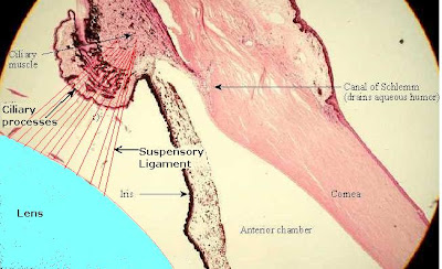
The ciliary processes are attached by their periphery to three or four of the ridges of the orbiculus ciliaris, and are continuous with the layers of the choroid. Their opposite extremities are free and rounded, and are pointing towards the posterior chamber of the eyeball and circumference of the lens.

Ciliary Body

The ciliary body is the circumferential tissue inside the eye which is made up of the ciliary muscle and ciliary processes. It is formed by the inward folding of the various layers of the choroid. The ciliary body is coated by a double layer of tissue, the ciliary epithelium. The inner layer is transparent and covers the vitreous body, and is continuous from the neural tissue of the retina. The outer layer is highly pigmented, continuous with the retinal pigment epithelium, and constitutes the cells of the dilator muscle.

The ciliary body releases a transparent liquid which is called the aqueous humor, which provides most of the nutrients for the lens and the cornea. The other function of the ciliary body is to change the shape of the lens to make the vision adapt for far sight or near sight.

Wednesday, March 18, 2009

Zonule of Zinn

The zonule of Zinn is a ring of fibrous strands which connects the ciliary body with the eye lens. These fibers are known as the suspensory ligament, which holds the lens in place. It relaxes by the contraction of the ciliary muscle. Relaxation of the ligament allows the lens to become more convex, adapting to short range focus.

The zonule is divided into two layers: a thin layer which lines the hyaloid fossa and a thicker layer which consists of zonular fibers. The zonule of Zinn is a system of fibers that stretches from the ciliary body to the lens periphery. Its function is to secure the lens in the optical axis and transfer forces from the ciliary muscle in accommodation.

Tuesday, March 17, 2009

Ciliary Muscle

The ciliary muscle is the muscle that changes the shape of the eye lens to accomodate the sight for distant or near vision. The ciliary muscle is connected to suspensory ligaments called zonule of Zinn or ciliary zonule.

When the ciliary muscle contracts, it releases the tension on the lens caused by the suspensory ligaments. This release of tension makes the lens more spherical, adapting to short range vision. When the ciliary muscle relaxes the ligaments are taut, stretching the lens thin enabling it to focus on distant objects.

The autonomic nervous system controls the contraction and relaxation of the ciliary muscle. Sympathetic nerve fiber stimulation is responsible for muscle relaxation, whereas parasympathetic stimulation causes muscle contraction. The ciliary muscle is an extension of the ciliary body.


Monday, March 16, 2009

Human Eye

The human eye is a sense organ that allows humans conscious light perception, vision, and color. The human eye consists of a pair of eyeballs set in the skull sockets, or orbital bone, below the brow.

The eyeball is made up of three layers of tissue:

1) Sclera, which an external tough layer made of fibrous tissue, wraps up the eye, except at the front where it becomes thin and transparent to form the cornea. The sclera is commonly referred to as the white of the eye.

2) Choroid is the middle vascular layer which lies between the sclera and the retina. It ends at the front with the ciliary processes and the ciliary muscle, which holds the lens of the eye.

3) The retina is a the inner layer of the eyeball. It consists of light, sensitive tissue that lines the choroid. It is on the retina where all images that we see are focused on and transmitted through the optic nerve to the brain.

The cornea is at the front of the eyeball which is a fine transparent extension of the sclera. The iris is located also at the front, but beyond the cornea and it consists of a layer of pigmented muscles, which are composed of two types of fibers, radial fibers and circular fibers. These fibers open and close the pupil when they contract.

The lens is set just behind the iris, blocking the pupilar orifice, and held in place by the ciliary muscles. Between the cornea and the lens there is aqueous humor, which a liquid substance. The inner hollow of the eyeball is filled with the vitreous humor, a transparent, jelly-like liquid.

The images of the objects that we see are refracted by the cornea and then is focused on the retina by the lens, whose shape is altered by the ciliary muscle to make the vision adapt to look at objects situated at different distances.

The eyeball movement is controlled by six types of external muscles attached to the sclera and anchored to the orbital bones of the eye sockets. They are the medial rectus muscle, superior rectus muscle, inferior rectus muscle, lateral rectus muscle, inferior oblique muscle, and superior oblique muscle.


Saturday, March 14, 2009

Proton

The proton is a positively charged subatomic particle which is located in the nucleus of each atom. It consists of 3 fundamental particles, two up quarks and one down quark. Protons and neutrons are nucleons, that may be bound by the nuclear force into atomic nuclei. The nucleus of the most common isotope of the hydrogen atom is a single proton.


Friday, March 13, 2009

Pion

A pion is the lightest of the meson family of elementary particles. A pion is a multiplet of three particles. The neutral pion has a mass about 264 times that of the electron. The charged pions, p+ and p-, have a mass about 273 times that of the electron. The neutral pion is its own antiparticle, and the negative pion is the antiparticle of the positive pion. Each pion is made up of a quark bound to an antiquark. Free pions are unstable. Charged pions decay with an average lifetime of 2.55 × 10⁻⁸ sec into a muon of like charge and a neutrino or antineutrino, while the neutral pion decays in about 10-15 sec, usually into a pair of photons but occasionally into a positron-electron pair and a photon.

Pions have zero spin and consist of first-generation quarks. In the quark model, an up and an anti-down quark compose a p+, while a down and an anti-up quark compose the p-, its antiparticle. The neutral combinations of up with anti-up and down with anti-down have identical quantum numbers, so they are only found in superpositions. The lowest-energy superposition is the p0, which is its own antiparticle. Together, the pions form a triplet of isospin; each pion has isospin-1 (I = 1) and third-component isospin equal to its charge (Iz = +1, 0 or -1).

The existence of the pion was predicted in 1935 by Hideki Yukawa, who theorized that it was responsible for the force of the strong interactions holding the atomic nucleus together. It was first detected in cosmic rays by C. F. Powell in 1947.

Mesons

Mesons are subatomic particles which are composed of one quark and one antiquark. They belong to the hadron particle family. The other members of the hadron family are the baryons, which are subatomic particles composed of three quarks. The main difference between mesons and baryons is that mesons are bosons while baryons are fermions. That is to say that mesons have integer spin while baryons have half-integer spin, which means that the Pauli exclusion principle does not apply to them.

Mesons take part in both the weak and strong interactions. Mesons with net electric charge also participate in the electromagnetic interaction. They are classified depending on their quark content, total angular momentum, parity, and various other properties such as C-parity and G-parity. Each meson has a corresponding antiparticle which is called antimeson.

Thursday, March 12, 2009

Baryons

Baryons are composite particles made of up three quarks. baryons and mesons are part of the larger particle family which comprises all particles made up of quarks; the hadrons. The term baryon is derived from the Greek barys, which means "heavy", because at the time of their naming it was believed that baryons were characterized by having greater masses than other particles.

As baryons are composed of quarks, they take part in the strong interaction. The most famous baryons are the protons and neutrons which constitute most of the mass of the visible matter in the universe, whereas electrons, the other major component of atoms, are leptons. Each baryon has a corresponding antiparticle where quarks are replaced by their corresponding antiquarks. For example, a proton is made of two up quarks and one down quark. Its corresponding antiparticle, the antiproton, is made of two up antiquarks and one down antiquark.

Baryons are strongly interacting fermions, which means that they experience the strong nuclear force and are described by Fermi-Dirac statistics, which apply to all particles obeying the Pauli exclusion principle. The bosons, on the other hand, do not obey the exclusion principle.

Baryons, together with mesons, are hadrons. That is to say they are particles composed of quarks. Quarks have baryon numbers of B = 1/3 and antiquarks have baryon number of B = -1/3. The term "baryon" usually refers to triquarks—baryons made of three quarks (B = 1/3 + 1/3 + 1/3 = 1).


Wednesday, March 11, 2009

Hadrons

Hadrons are particles which are held together by their strong interaction. Hadrons include mesons and baryons but excludes leptons, which do not interact by the strong force. The weak interaction acts on both hadrons and leptons.

Hadrons consist of quarks, either as quark-antiquark pairs (mesons) or as three quarks (baryons). Quarks are surrounded by a cloud of gluons, the exchange particles for the color force. Recent experimental evidence has showed the existence of five-quark combinations that are being called pentaquarks.

Hadrons are assigned quantum numbers which correspond to the representations of the Poincaré group: JPC(m), where J is the spin quantum number, P, the intrinsic (or P) parity, and C, the charge conjugation, or C parity, and the particle four-momentum, m.

Tuesday, March 10, 2009

Quarks

Quarks are matter particles which constitute neutrons and protons. They are the only particles in to experience all four fundamental forces, which are also known as fundamental interactions. There are six different types of quarks. Each quark type is called a flavor. Single quarks are not usually found on their own. They can only be found in composite particles called hadrons, such as protons and neutrons. Thus much of what we know about quarks has been inferred from observations on the hadrons themselves.

As they are confined by the strong force fields, quarks only exist inside hadrons. As a result, it is impossible measure their mass by isolating them. There are six types of quarks which are known as flavors: up (u), down (d), charm (c), strange (s), top (t) and bottom (b). The up and down quarks have the lowest masses of all quarks, and thus are generally stable and very common in the universe. The other quarks are much more massive, and will rapidly decay into the lighter up and down quarks. The heavier charm, strange, top and bottom quarks can only be produced in high energy collisions, such as in particle accelerators and cosmic rays.

Tau Lepton

The tau lepton is a negatively charged, electron-like particle with a mass of 1.784 GeV/c2. It has a a mass of 1,777 MeV/c2. Its antiparticle is the tau-plus, which has the same mass but a positive electric charge. The tau lepton particles were discovered at SLAC in experiments at SPEAR. The 1995 Nobel Prize was awarded to Martin L. Perl from Stanford University, CA, USA, for this discovery.

Monday, March 9, 2009

Giant Magnetoresistance

Giant magnetoresistance is the change in electrical resistance of some materials in response to an applied magnetic field. The application of a magnetic field to magnetic metallic multilayers such as Fe/Cr and Co/Cu, in which ferromagnetic layers are separated by nonmagnetic spacer layers of a few nm thick, results in a significant reduction of the electrical resistance of the multilayer. This effect was found to be much larger than other magnetoresistive effects that had ever been observed in metals and was, therefore, called “giant magnetoresistance”. In Fe/Cr and Co/Cu multilayers the magnitude of GMR can be higher than 100% at low temperatures.

Giant Magnetoresistance is a very large change in electrical resistance that is observed in a ferromagnet/paramagnet multilayer structure. Resistance change occurs when the relative orientations of the magnetic moments in alternate ferromagnetic layers change as a function of applied field. In the absence of the magnetic field the magnetizations of the ferromagnetic layers are antiparallel. Applying the magnetic field, which aligns the magnetic moments and saturates the magnetization of the multilayer, leads to a drop in the electrical resistance of the multilayer.

Friday, March 6, 2009

Positron

The positron is the antiparticle of the electron. The positron has the same mass as an electron, an electric charge of +1, and a spin of 1/2. When a low-energy positron collides with a low-energy electron, there is mutual annihilation, which results in the production of two gamma ray photons. Positrons can be generated by positron emission radioactive decay, or by pair production from a sufficiently energetic photon. The positrón was discovered by Carl David Anderson in 1932 when he studied the cosmic ray.

Quantum Mechanics

Quantum mechanics is a set of principles which underlies fundamental known description of all physical systems at the microscopic scale at the atomic level. It is the study of matter and radiation at an atomic level. Among these principles are both a dual wave-like and particle-like behavior of matter and radiation, and prediction of probabilities in situations where classical physics predicts certainties. Classical physics can be derived as a good approximation to quantum physics, typically in circumstances with large numbers of particles. Thus quantum phenomena are relevant in systems whose dimensions are close to the atomic scale, such as molecules, atoms, electrons, protons and other subatomic particles.

The word quantum refers to a discrete unit that quantum theory assigns to certain physical quantities, such as the energy of an atom at rest. Quantum mechanics is vital in order to comprehend the behavior of systems at atomic length scales. If classical mechanics governed the workings of an atom, electrons would rapidly travel towards and collide with the nucleus, making stable atoms impossible. Nevertheless, in the natural world the electrons usually remain in an unknown orbital path around the nucleus, defying classical electromagnetism.

Thursday, March 5, 2009

Spontaneous Broken Symmetry

Spontaneous broken symmetry occurs when a system possessing a certain symmetric property collapses into a vacuum state which does not possess the symmetric property.

To help explain spontaneous broke symmetry a common example is usually given; a ball sitting on top of a hill in a completely symmetric state. However, its state is unstable: the slightest disturbance will cause the ball to roll down the hill in some particular direction into its lowest energy state. At that point, symmetry has been broken. A symmetrical situation therefore collapses into an asymmetrical state.

Spontaneous broken symmetry conceals nature’s order under a seemingly jumbled surface. In 1960, Yoichiro Nambu formulated the mathematical description of spontaneous broken symmetry in elementary particle physics. The spontaneous broken symmetries which Nambu formulated, differ from the broken symmetries described by Toshihide Maskawa and Makoto Kobayashi. These spontaneous occurrences seem to have existed in nature since the very beginning of the universe and came as a complete surprise when they first appeared in particle experiments in 1964.

Wednesday, March 4, 2009

Antibiotic

An antibiotic is medicine that fights bacterial infections, by killing or inhibiting the growth of bacteria. There are two types of antibiotics; bactericidal and bacteriostatic. A bactericidal antibiotic destroys bacteria, and a bacteriostatic inhibits the growth of bacteria by interfering with bacterial protein production, DNA replication, or other aspects of bacterial cellular metabolism.Used properly, antibiotics can save lives.

Anti-bacterial antibiotics are categorized based on their target specificity: narrow-spectrum antibiotics target particular types of bacteria, such as Gram-negative or Gram-positive bacteria, while broad-spectrum antibiotics affect a wide range of bacteria.

There are several classes of antibiotics:
1) Aminoglycosides: gentamicine, amikacin, neomicyn, streptomycin, etc. Efective in the treatment against infections caused by Gram-negative bacteria, such as Escherichia coli and Klebsiella particularly Pseudomonas aeruginosa. Effective against Aerobic bacteria.
2) Cephalosporins: Cefadroxil, Cefazolin, etc.
3) Macrolides: azithromycin, clarithromicyn, erythromicyn, etc. They are used in the treatment of streptococcal infections, syphilis, respiratory infections, mycoplasmal infections, Lyme disease.
4) Penicillins: amoxicillin, ampicillin, penicillin, oxacillin, etc. Used against a wide range of infections; penicillin is used against streptococcal infections, syphilis, and Lyme disease.

Tuesday, March 3, 2009

Amikacin

Amikacin is a bactericidal antibiotic used to treat different types of bacterial infections. Amikacin belongs to the aminoglycoside group of antibiotics and works by binding to the bacterial 30S ribosomal subunit, causing misreading of mRNA and leaving the bacterium unable to synthesize proteins vital to its growth.

Amikacin is used for treating infections of central nervous system, urogenital system, biliary and intestinal tracts, intraabdominal infections, and pneumonia, caused by Gram-negative microorganisms, secondary infections after combustion, bacterial septicemia, infections of the bones and joints.

Amikacin is administered once or twice a day by the intravenous or intramuscular route, which tends to be painful. There is no oral form available.

Monday, March 2, 2009

Streptomycin

Streptomycin is a bactericidal antibiotic which belongs to aminoglycoside group of antibiotics. It was the first to be discovered and the first remedy for tuberculosis. Streptomycin is derived from the actinobacterium Streptomyces griseus. It kills sensitive microbes by inhibiting protein synthesis. More specifically, it binds to the 16S rRNA of the bacterial ribosome and interferes with the binding of formyl-methionyl-tRNA to the 30S subunit.

Streptomycin is a water-soluble aminoglycoside. The chemical name of streptomycin sulfate is D-Streptamine.